Antibodies and FC fusion protein modifications with enhanced persistence or pharmacokinetic stability in vivo and methods of use thereof
a technology of fusion polypeptides and antibodies, which is applied in the field of enhancing the in vivo persistence and stability of can solve the problems of mutagenesis and testing, difficulty in synthesizing long monodisperse species, and difficulty in mutagenesis and testing, so as to increase the half-life of circulating antibodies and fc fusion polypeptides and increase the serum half-life
- Summary
- Abstract
- Description
- Claims
- Application Information
AI Technical Summary
Benefits of technology
Problems solved by technology
Method used
Image
Examples
example 1
Biotinylation of Human Immunoglobulin G
[0141]Purified human immunoglobulin G (hIgG) (Gammagard, Baxter) was conjugated with biotin by first diluting the hIgG stock in 50 mM sodium phosphate buffer, pH 7.2 with the same buffer to 2.2 mg / ml, or 1.37×10⁻⁵ M. N-hydroxysuccinimidobiotin (EZ-Link™ NHS-biotin, Pierce cat. #20217) was allowed to come to room temperature, then a 3.42×10⁻³ M solution of NHS-biotin in DMSO (10× strength) was produced. One volume of 3.42×10⁻³ M (10×) NHS-biotin was added to 9 volumes of 1.37×10⁻⁵ M hIgG to achieve a 25-fold excess molar concentration of NHS-biotin. The reaction was performed for 30 minutes at room temperature, then 1 volume of 1 M glycine, pH 7.2 was added to stop the reaction. The reactants were twice dialyzed in 5 liters of 50 mM sodium phosphate buffer, pH 7.2 at 4° C. The hIgG-biotin solution was sterile filtered using a 0.45 μm pore syringe filter and stored at 4° C. To determine the biotin:hIgG ratio the EZ Biotin Quantitation Kit (Pierce...
example 2
Determination of hIgG-biotin Half Life In Vivo
[0143]To determine the effect of biotinylation on the in vivo half life of hIgG, C57BL / 6 mFcRn− / − hFcRn + / Tg Line 276 mice (3-5 mice / group) were given intraperitoneal injections of 100 μg of hIgG with, or without, biotin conjugation at day-3. C57BL / 6 mFcRn− / − hFcRn + / Tg Line 276 mice do lack the mouse FcRn gene and do express the human FcRn. For a more detailed description of the mouse model, see (Petkova et al., 2006a). Plasma was obtained 3 days after tracer injection (to avoid the alpha phase of hIgG degradation), at day 0, 7, 14, 21, and 28, by collecting either 25 μl, or 75 μl of blood from the retro-orbital sinus into capillary tubes and transferring to 1.5 ml microcentrifuge tubes containing 2 μl of PBS with 10,000 U / ml heparin. Tubes were centrifuged at 10,000 rpm for 5 minutes at 4° C. in an Eppendorf 5417R centrifuge (New York). Plasma samples were transferred to 96 well round bottom polypropylene storage plates (Corning cat. #...
example 3
Correlation of Degree of Biotinylation to Plasma Half-life
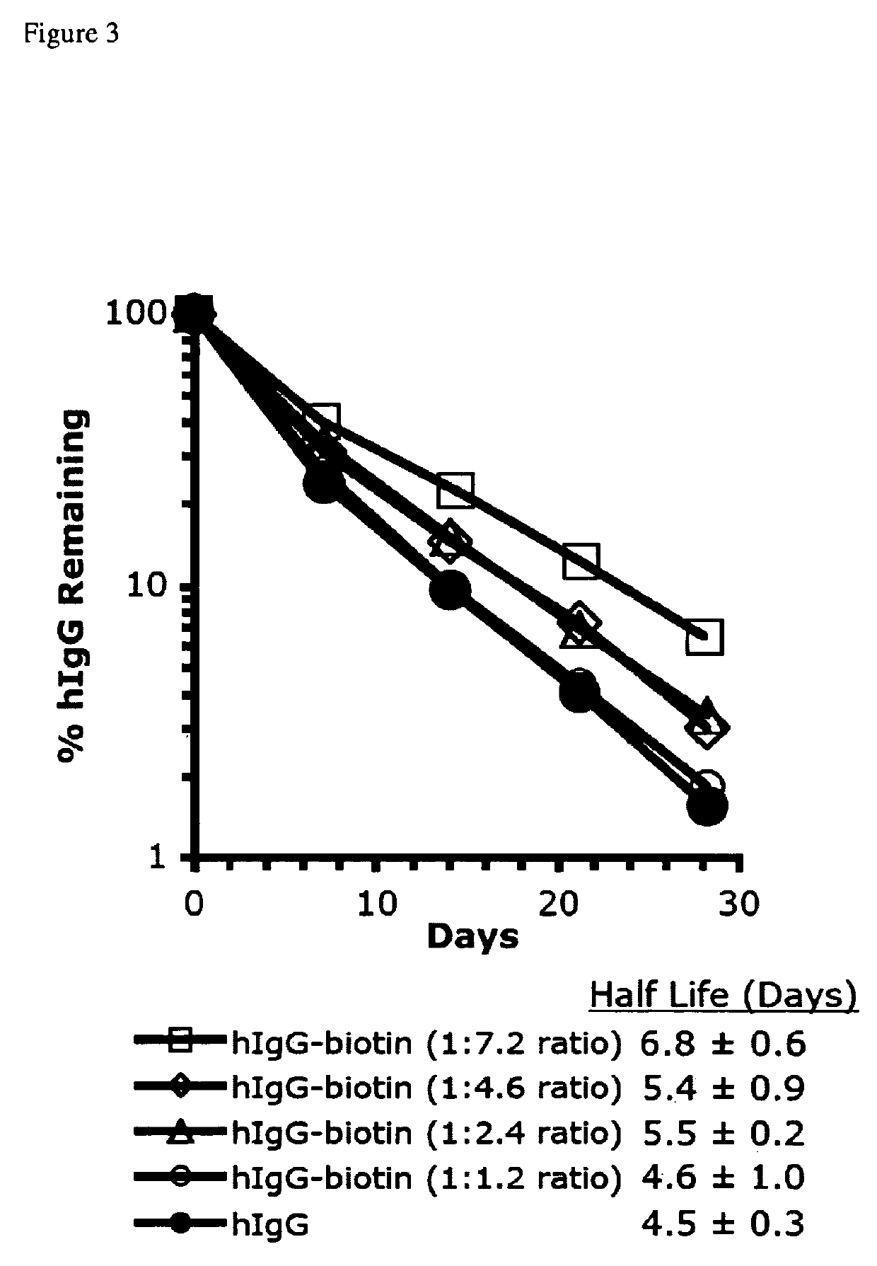
[0149]To determine what degree of biotin conjugation was necessary to effect an increased half life, hIgG was labeled as described in Example 1, but with the reaction performed on ice to slow the rate to allow more precise labeling ratios, and the length of reaction was varied to obtain the biotin:hIgG ratios 1.2, 2.4, 4.6, and 7.2. As a model to analyze the plasma half life in vivo, we have used a mouse model expressing the human FcRn while lacking the mouse FcRn receptor on the C57BL / 6 background (C57BL / 6 FcRn− / −; hFcRn Tg276), which is described in (Petkova et al., 2006a). 100 μg of each of these hIgG-biotin tracers and hIgG were intraperitoneally injected into C57BL / 6 mFcRn− / − hFcRn + / Tg Line 276 mice (5 mice / group) at day-3. Plasma was collected from mice on day 0, 7, 14, 21, and 28, and measured by hIgG ELISA as described in Example 2.
[0150]The conjugation of biotin to available lysine residues on hIgG increased this tr...
PUM
| Property | Measurement | Unit |
|---|---|---|
| molar ratio | aaaaa | aaaaa |
| pH | aaaaa | aaaaa |
| molar concentration | aaaaa | aaaaa |
Abstract
Description
Claims
Application Information
 - R&D
- Intellectual Property
- Life Sciences
- Materials
- Tech Scout
- Unparalleled Data Quality
- Higher Quality Content
- 60% Fewer Hallucinations
Browse by: Latest US Patents, China's latest patents, Technical Efficacy Thesaurus, Application Domain, Technology Topic, Popular Technical Reports.
© 2025 PatSnap. All rights reserved.Legal|Privacy policy|Modern Slavery Act Transparency Statement|Sitemap|About US| Contact US: help@patsnap.com


