Method for isolating hepatocytes
A technology of hepatocytes and cells, applied in the field of separation of hepatocytes, can solve problems such as rejection reaction
- Summary
- Abstract
- Description
- Claims
- Application Information
AI Technical Summary
Problems solved by technology
Method used
Image
Examples
Embodiment 1
[0058] Example 1: Collection of hepatocytes after hepatectomy
[0059] Hepatocytes from 5 patients who underwent liver resection due to liver metastases. The study was approved by the Ethics Committee of St George's Hospital, New South Wales, Australia (approval number 01 / 123). Details of the localization and resection of metastatic tumors in these patients are shown in Table 1.
[0060] patient
(gender)
liver resection day
Expect
resected segment
(cm)
1(female)
CRC 2 -Mar 01
April 2003
4
4×3×2
2(male)
CRC-Nov 00
May 2002
2, 3 & 4
(Collect 2+
3)
4×4×2
3 (female)
CRC-Nov 00
April 2002
2, 3
2×2×1.5
4(male)
CRC-Apr 01
May 2002
2, 3 & 7
(Collect 2+
3)
2×2.5×2
4.5×2.7×
2
5(male)
Pancreatic Cancer - Apr 01
May 2002
5, 6 ...
Embodiment 2
[0066] Example 2: Isolation of tumor-free hepatocytes
[0067] After obtaining viable hepatocytes (Example 1), the hepatocytes were isolated from the accompanying tumor cells. Tumor cells were isolated by the immunomagnetic separation method described by Flatmark et al. (Clinical Cancer Research 8:444-449, 2002) using 4.5 μm superparamagnetic beads (Dynabeads M450; Dynal, Oslo, Norway) coated with MOC31 monoclonal antibody. MOC31 recognizes the Ep-CAM antigen, which is present on the surface of most epithelial cells and is particularly highly expressed in colorectal cancer.
[0068] 5 million hepatocytes were mixed with 1 million HT29 colorectal cells in 1 ml of phosphate buffered saline (PBS). 200 μl of Dynabeads M450 were suspended in 1 ml of PBS, and 20 μl of MOC31 antibody was added. The suspension was incubated at 4°C for 30 minutes before adding the MOC31-coated Dynabeads mixture to the tube containing the hepatocytes plus HT29 cell mixture to a total volume of 2 ml. ...
Embodiment 3
[0069] Example 3: Validation of Isolation of Tumor-Free Hepatocytes
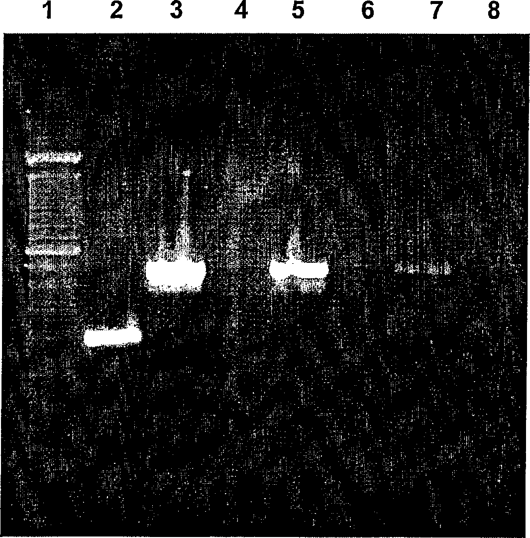
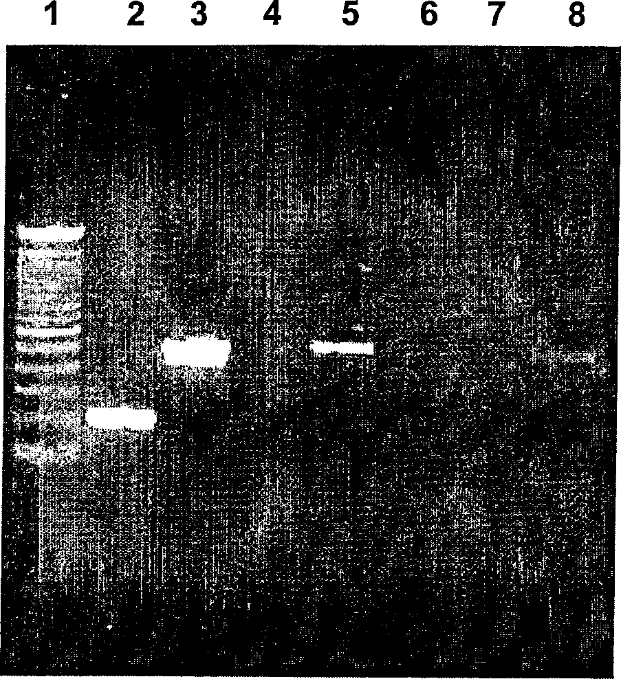
[0070] After tumor cells were removed using MOC31-coated immunomagnetic beads (Example 2), the prepared hepatocytes were analyzed for residual tumor cells by detecting the expression of the epithelial cell adhesion molecule (Ep-CAM) gene using RT-PCR . Ep-CAM is a useful cell surface marker expressed on the surface of most epithelial and tumor cells including HT29. The sensitivity of RT-PCR in tumor cell detection based on Ep-CAM gene expression is about 10 tumor cells / 10 7 non-tumor cells (Sakaguchi, M et al., Brit J Cancer 79:416-422, 1999).
[0071] The following primers (primer) were used for RT-PCR analysis:
[0072] Sense strand: 5'-GAACAATGATGGGCTTTATGA-3'
[0073] Nonsense strand (antisense strand): 5'-TGAGAATTCAGGTGCTTTTT-3'
[0074] Successful PCR amplification of Ep-CAM using these primers generated a 515 bp product.
[0075] Hepatocytes were obtained as described in Example 1. Then, hepato...
PUM
 Abstract
Description
Claims
Application Information
 - R&D
- Intellectual Property
- Life Sciences
- Materials
- Tech Scout
- Unparalleled Data Quality
- Higher Quality Content
- 60% Fewer Hallucinations
Browse by: Latest US Patents, China's latest patents, Technical Efficacy Thesaurus, Application Domain, Technology Topic, Popular Technical Reports.
© 2025 PatSnap. All rights reserved.Legal|Privacy policy|Modern Slavery Act Transparency Statement|Sitemap|About US| Contact US: help@patsnap.com


