
Stable-state front mechanical transverse wave imaging method for magnet resonance elastic diagram technology

A technology of magnetic resonance elasticity and imaging method, which is applied in the field of imaging in the field of bioengineering technology, can solve different problems and achieve the effect of avoiding reflection

Inactive Publication Date: 2006-08-09
SHANGHAI JIAO TONG UNIV
View PDF0 Cites 5 Cited by
  • Summary
  • Abstract
  • Description
  • Claims
  • Application Information

AI Technical Summary

Problems solved by technology

It differs from existing steady-state fluctuation imaging methods

Method used


Image

  • Stable-state front mechanical transverse wave imaging method for magnet resonance elastic diagram technology
  • Stable-state front mechanical transverse wave imaging method for magnet resonance elastic diagram technology

Examples


Embodiment Construction

[0024] In order to better understand the technical solutions of the present invention, a further detailed description will be made below in conjunction with the accompanying drawings and implementation examples.

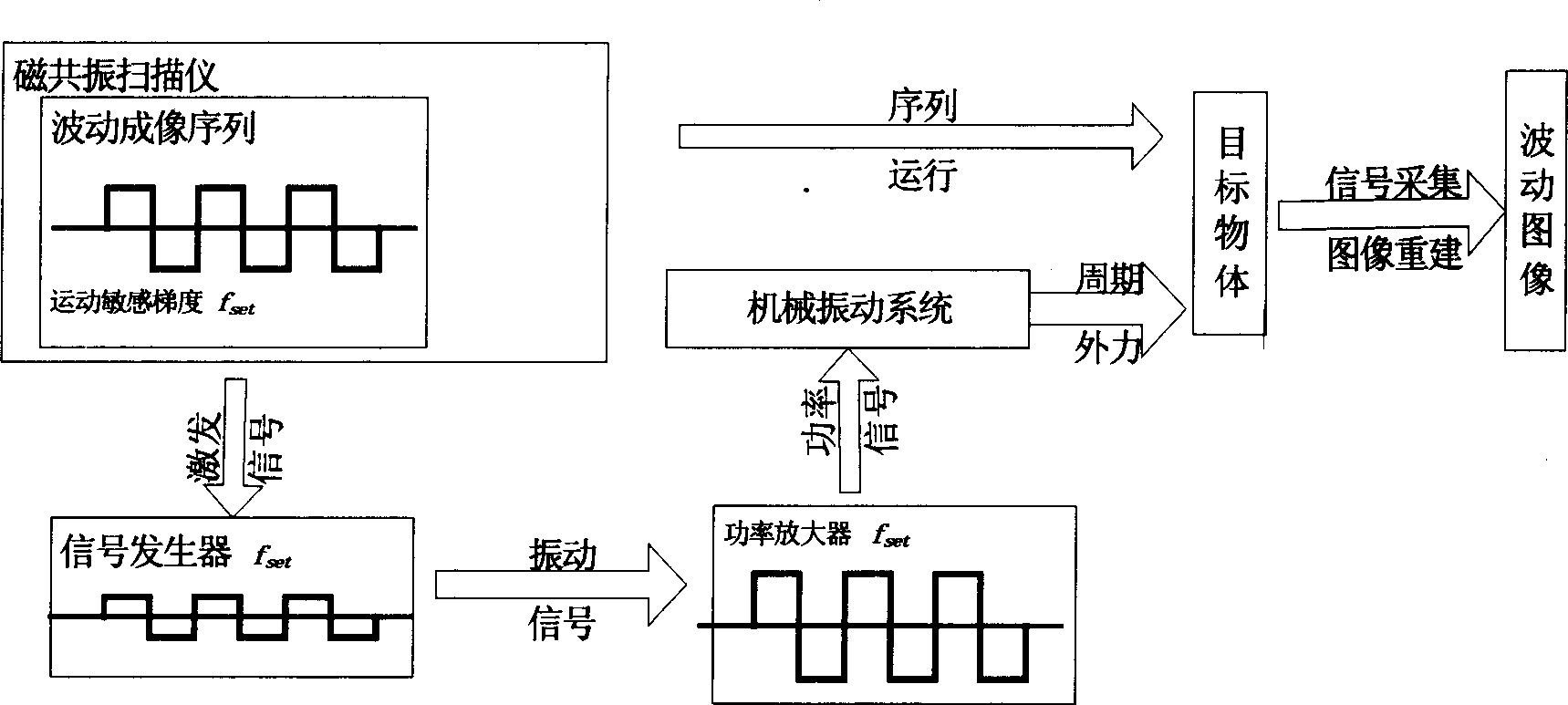
[0025] The invention takes the agarose gel imitation body as the imaging object, and implements the mechanical shear wave imaging before the steady state. Such as figure 1 Shown is a schematic diagram of the system of the present invention. The whole imaging system includes wave imaging sequence (including motion-sensitive gradient square wave), magnetic resonance scanner, signal generator, power amplifier, and mechanical vibration system. Before imaging, first set up the system, connect the mechanical vibration system, power amplifier, signal generator and magnetic resonance scanning instrument, connect the prepared cylindrical agarose phantom with the mechanical vibration system, and put them into the magnetic resonance scanner in the magnet cavity.

[0026] Aft...


PUM


Abstract

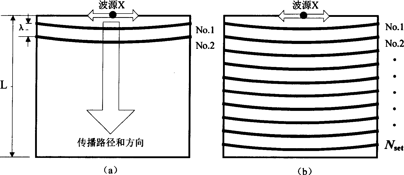
The present invention relates to a mechanical transverse wave imaging method before steady-state in the magnetic resonance elastic image technology, belonging to the field of bio-engineering technology. Said invention is characterized by that when the MRE image-formation is made, the imaging time can be controlled, so that before the moment of that the mechanical transverse wave is reached to tissue boundary, the wave image-formation is implemented, in which the time of that the mechanical transverse wave is reached to boundary can be obtained by utilizing anatomical structure size of tissue and wave speed calculation obtained by steady-state wave image-formation, on this basis, according to the set mechanical transverse wave frequency the number of cycles of the mechanical vibration and number of kinetic sensitive gradient pulses in the pulse sequence can be calculated so as to set the wave imaging sequence parameter before steady-state and make the mechanical transverse wave be imaged.

Description

technical field [0001] The present invention relates to an imaging method in the technical field of bioengineering, in particular to a steady-state pre-mechanical shear wave imaging method in magnetic resonance elastography. Background technique [0002] Magnetic resonance elastography (MRE) is a medical imaging technique currently under development that can non-invasively measure the biomechanical properties of biological tissues. Although people have studied the mechanical properties of materials for hundreds of years, it is difficult to obtain the biomechanical parameters of living biological tissues. The main reason is that the mechanical load test for measuring the mechanical properties of materials in the laboratory is difficult to apply in the internal tissues of living organisms. Magnetic resonance elastography is used to measure the distribution of elastic coefficients inside living biological tissues. It first uses a vibrating device to excite the surface of the t...

Claims


Application Information

Patent Timeline
IPC IPC(8): A61B5/055G01R33/20G01N24/00
Inventor 陈俊缪飞庄天戈
Owner SHANGHAI JIAO TONG UNIV
Learn More