Method for fabricating section of biologic tissue

A production method and biological tissue technology, which are applied in the preparation of test samples, microscopy, optics, etc., can solve the problems of inability to obtain an ideal section of tissue specimens, unfavorable excavation, and folded boundary lines. Straightforward interface

Inactive Publication Date: 2005-10-26
SHANTOU UNIV MEDICAL COLLEGE
View PDF0 Cites 5 Cited by
  • Summary
  • Abstract
  • Description
  • Claims
  • Application Information

AI Technical Summary

Problems solved by technology

[0003] However, when studying flat and layered tissue specimens (such as skin or mucous membranes) in medicine, it is often desired that the cut plane be perpendicular to the interface between the various layers of the specimen, so that the interface between the various layers of the obtained slice is straight, especially It is hoped that the target cells or target tissue layers in the slice can be easily and quickly dug out under the microscope. In this case, the shortcomings of the traditional biological tissue slice production method will appear, because: 1. If the tissue specimen is flattened And tiled on the stage, the flat tissue specimen stretching direction will be basically parallel to the direction of the stage’s back and forth movement, the obtained section will be basically parallel to the interface between the layers of the tissue sample, and the obtained section will be in the Theoretically, it is likely to be only a certain layer of the tissue sample, and the ideal section perpendicular to the interface between the layers of the tissue sample cannot be obtained; The boundary between layers of tissue will be wrinkled and not straight, and it is not conducive to easy and straightforward digging out of the target cells or target tissue layers in the slice under the microscope

Method used


Image

  • Method for fabricating section of biologic tissue
  • Method for fabricating section of biologic tissue
  • Method for fabricating section of biologic tissue

Examples


Embodiment 1

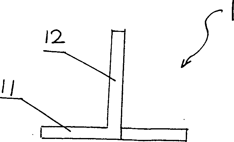
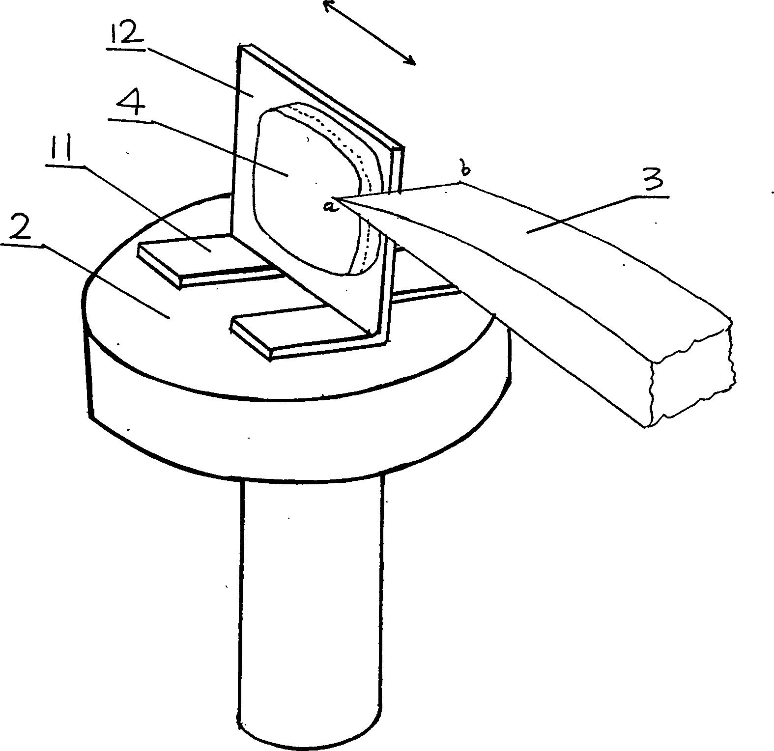
[0024] This method is applied to make a flat shape and a three-layer structure (i.e. Figure 4 41 layers, 42 layers, 43 layers) of the tissue specimen slices, including the following main steps: A, take out the flat tissue specimen 4 with a layered structure, and use its own adhesive force to initially adhere it to an inverted T-shaped plastic bracket 1 on the vertical plate 12, and stretched and flattened; the bottom plate 11 of the inverted T-shaped plastic bracket is laid flat and glued on the stage 2 of the microtome, so that the vertical plate 12 of the plastic bracket is positioned at the front of the blade of the knife 3. In the front, make the plane where the vertical plate 12 of the plastic support is located and the direction of movement back and forth when the stage 3 is working (i.e. image 3 The direction of the middle arrow) is basically parallel, such as image 3 As shown; the flat tissue specimen 4 is frozen, and it is further frozen and fixed on the vertical ...

Embodiment 2

[0026] The method comprises the following major steps: A, flattening the flat tissue specimen with layered structure on the vertical plate of the inverted T-shaped plexiglass support, embedding with paraffin, making the bottom plate of the plexiglass support flat and fixed on the rotary On the stage of the cutting microtome, make the vertical plate of the plexiglass support stand in front of the knife blade, and make a line between the plane where the vertical plate of the plexiglass support is located and the starting point and end point of each back and forth movement of the stage The directions intersect to form an included angle of about 30°; B. Drive the movement of the stage of the microtome, the stage drives the plexiglass bracket and the tissue specimen to move relative to the knife, and the knife cuts the tissue specimen together with the vertical plate of the plastic bracket step by step Slicing; C. Put the slices obtained in step B into hot water, separate the tissue...


PUM


Abstract

The invention is the biological tissue slice making method, which comprises the following processes: A. flatten the tissue sample and fix on the vertical plate of the inverse T-shape non-metal bracket, and fix the base plate of the bracket on the carrying platform and make the vertical plate of the bracket in front of the knifeware and parallel the platform of the bracket and the moving direction of the carrying platform; B. the slicing machine simultaneously slices the tissue samples and the vertical plate of the non-mental bracket. The invention makes the slicing face vertical to the interface of each tissue sample layer, and every interface is flat and straight.

Description

technical field [0001] The invention relates to a method for making biological tissue slices. Background technique [0002] When the existing biological tissue slices are made, the biological tissue specimens are fixed on the stage of the microtome. The fixing methods mainly include low-temperature freezing fixation or paraffin embedding; The plane on which the surface of the object stage is located is basically parallel, and the stage will automatically step a short distance after each round of back and forth motion, and the direction of the stepping motion is perpendicular to the direction of the back and forth motion, thus driving the tissue specimen to move back and forth relative to the stationary tool and stepping motion, the tissue specimen is cut into slices step by step. This method is suitable for the situation where the flatness of the interface between the obtained slices is not high, such as in the pathological diagnosis in the hospital, because the pathologica...

Claims


Application Information

Patent Timeline
IPC IPC(8): G01N1/06
Inventor 胡盛平
Owner SHANTOU UNIV MEDICAL COLLEGE
Why Eureka
Try Eureka