Zinc finger protein and its antibody preparation and use
A protein, antibody technology, applied in the direction of peptide/protein composition, application, antibody, etc.
- Summary
- Abstract
- Description
- Claims
- Application Information
AI Technical Summary
Problems solved by technology
Method used
Image
Examples
Embodiment 1
[0070] Example 1 Preparation of ZNF268 protein and preparation of anti-ZNF268 antibody
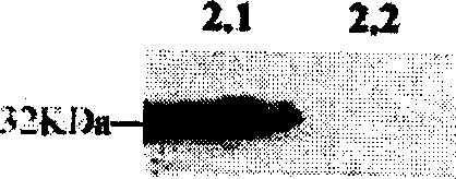
[0071] The 653bp fragment of ZNF268 was inserted into the prokaryotic expression vector pRSET-B to construct the recombinant prokaryotic expression vector pRSET-BH 2 , which encodes a ZNF268 protein fused to 6His at the N-terminus. pRSET-BH 2 The transformed E.coli BL21(DE3) was cultured to A 600 At 0.8, add 0.4mM IPTG and induce at 42°C for 3 hours, lyse the bacterial cells to obtain the crude cell extract containing soluble 6his-ZNF268 fusion protein, use XPress TM After the purification system was purified, Freund's adjuvant was added, and rabbits were immunized with standard methods to obtain anti-ZNF268 antibodies. figure 1 It shows that the ZNF268 protein is highly expressed under the experimental conditions. figure 2 It is shown in that the anti-ZNF268 antibody can specifically bind to the expressed ZNF268 protein.
Embodiment 2
[0072] Example 2 Protein hybridization analysis of ZNF268 protein expression
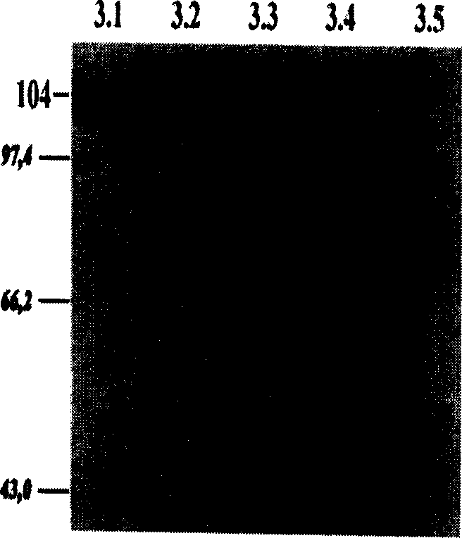
[0073] Total protein was extracted from different tissues of 4-month-old aborted human embryos with lysis buffer, separated by 8% SDS-PAGE and transferred to PVDF membrane. The membrane was blocked with TBST buffer containing 10% skimmed milk for 60 minutes, then incubated with TBST containing 1:640 diluted anti-ZNF268 antibody for 1 hour, and washed with TBST. Then the membrane was incubated in TBST containing appropriate goat anti-rabbit IgG antibody for 30 minutes. After TBST was eluted, the color reaction was carried out with DAB. image 3 It showed that ZNF268 was significantly expressed in fetal liver and intestine, but not expressed in fetal heart, kidney, brain and spleen.
Embodiment 3
[0074] Example 3 Immunohistochemical analysis of ZNF268 protein in the hematopoietic system
[0075] Different tissue materials were fixed with Bouin's solution, dehydrated with graded alcohol, transparent in xylene, and embedded in paraffin. The material was routinely sectioned and spread on glass slides previously treated with poly-L-lysine. Slides were blocked with normal goat serum and then reacted with anti-ZNF268 antibody. Biotin-conjugated anti-rabbit IgG was used as the secondary antibody, horseradish peroxidase-conjugated streptavidin was used as the third antibody, and DAB was used for color reaction. Finally, counterstaining was performed with hematoxylin. To identify cell types, monoclonal antibody anti-CD 34 , anti-CD 38 As the primary antibody, the corresponding biotin-conjugated anti-mouse IgG was used as the secondary antibody. PBS solution was used instead of the primary antibody as a control for non-specific staining of the experiment.
[0076] Figure...
PUM
 Abstract
Description
Claims
Application Information
 - Generate Ideas
- Intellectual Property
- Life Sciences
- Materials
- Tech Scout
- Unparalleled Data Quality
- Higher Quality Content
- 60% Fewer Hallucinations
Browse by: Latest US Patents, China's latest patents, Technical Efficacy Thesaurus, Application Domain, Technology Topic, Popular Technical Reports.
© 2025 PatSnap. All rights reserved.Legal|Privacy policy|Modern Slavery Act Transparency Statement|Sitemap|About US| Contact US: help@patsnap.com


