
Deep learning-based diseased lung CT segmentation and quantitative analysis method and system

A deep learning and quantitative analysis technology, applied in the field of pathological diagnosis, can solve the problems of difficult segmentation, adhesion of related soft tissues, and insignificant difference between the HU value of the lesion area and the lung contour, so as to reduce the difficulty of labeling and assist early screening. The effect of checking and diagnosing, reducing the amount of annotation

Pending Publication Date: 2022-07-29
THE FIRST AFFILIATED HOSPITAL ZHEJIANG UNIV COLLEGE OF MEDICINE
View PDF0 Cites 0 Cited by
  • Summary
  • Abstract
  • Description
  • Claims
  • Application Information

AI Technical Summary

Problems solved by technology

[0003] Traditional deep learning algorithms have a good segmentation effect on normal lung tissue or lungs with mild lesions, but in lung CT images with severe parenchymal lesions, because the difference between the HU value of the lesion area and the lung contour is not obvious, the relevant soft tissue Adhesive together, difficult to separate
Secondly, after the effective area of ​​the lungs is segmented, there is also a lack of evaluation of the air volume and lung weight of the lungs

Method used


Image

  • Deep learning-based diseased lung CT segmentation and quantitative analysis method and system
  • Deep learning-based diseased lung CT segmentation and quantitative analysis method and system
  • Deep learning-based diseased lung CT segmentation and quantitative analysis method and system

Examples


Embodiment Construction

[0038] The technical solutions in the embodiments of the present invention will be clearly and completely described below with reference to the accompanying drawings in the embodiments of the present invention. Obviously, the described embodiments are only a part of the embodiments of the present invention, rather than all the embodiments. Based on the embodiments of the present invention, all other embodiments obtained by those of ordinary skill in the art without creative efforts shall fall within the protection scope of the present invention.

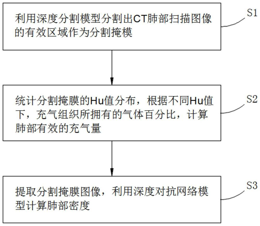
[0039] The present invention provides such as figure 1 Shown is a deep learning-based method for segmentation and quantitative analysis of diseased lung CT, the method includes the following steps:

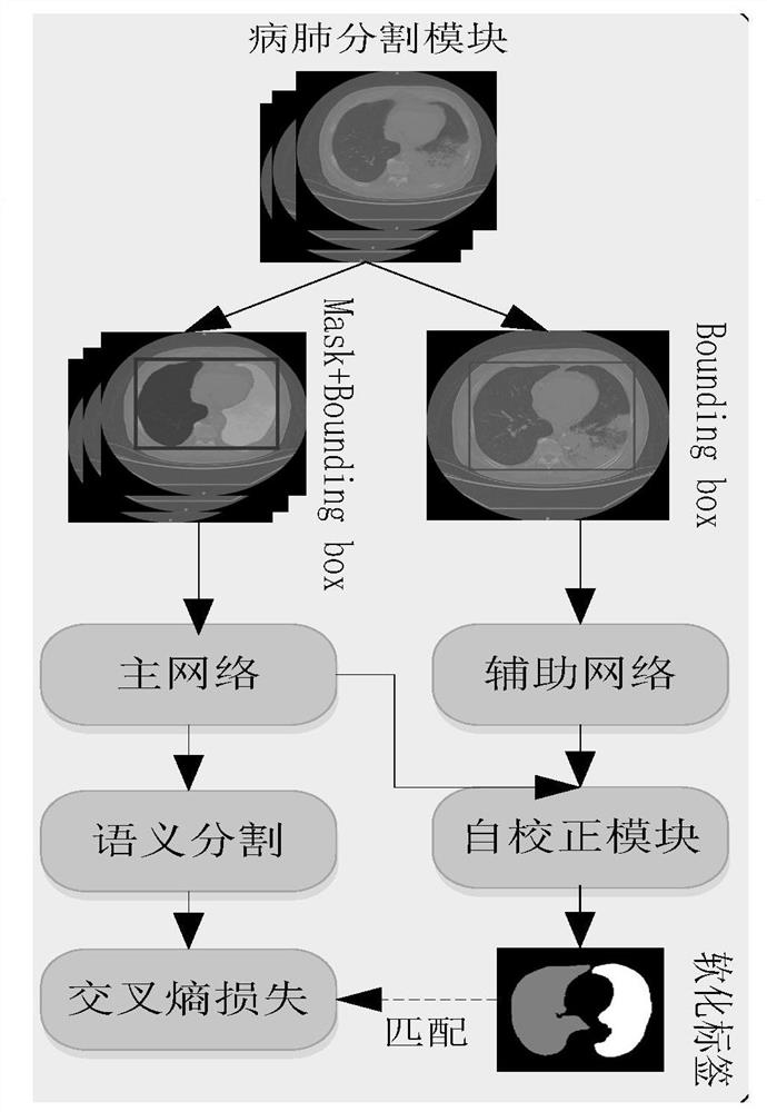
[0040] S1. Use a depth segmentation model to segment the effective area of ​​the CT lung scan image as a segmentation mask, and the depth segmentation model is trained by the main segmentation model, such as figure 2 As shown, the trai...


PUM


Abstract

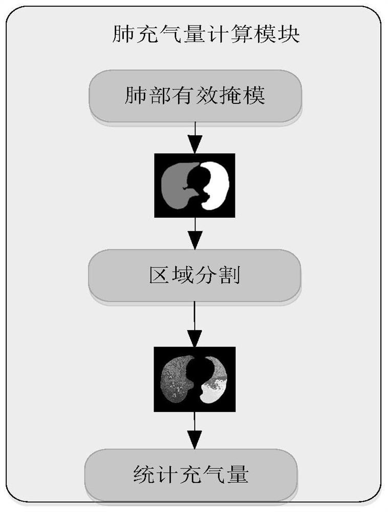
The invention discloses a disease lung CT segmentation and quantitative analysis method and system based on deep learning, and belongs to the technical field of pathological diagnosis, and the method comprises the following steps: segmenting an effective region of a CT lung scanning image through a deep segmentation model, and taking the effective region as a segmentation mask; carrying out statistics on Hu value distribution of the segmentation mask, and calculating the effective inflation volume of the lung according to the gas percentage of the inflated tissue under different Hu values; and extracting a segmented mask image, and calculating the lung density by using a deep adversarial network model. By means of the deep segmentation model, the labeling amount and labeling difficulty of data are greatly reduced, the lung inflation amount and the lung weight are calculated on the basis after the effective area of the lung is segmented, the general situation and development of the illness state of a patient can be observed in a multi-dimensional mode through the indexes, and a doctor can be assisted in early screening and diagnosis of the illness.

Description

technical field [0001] The invention belongs to the technical field of pathological diagnosis, and in particular relates to a method and system for CT segmentation and quantitative analysis of diseased lungs based on deep learning. Background technique [0002] In the segmentation and quantitative analysis of lung CT images, especially in the research of lung CT images with substantial lesions, the progress is slow. Accurate segmentation of the effective area of ​​the diseased lung is a basic step in lung research. The evaluation of effective lung air volume and lung weight has important significance for clinical diagnosis. [0003] The traditional deep learning algorithm has a good effect on normal lung tissue or lungs with mild lesions, but in the lung CT images with severe parenchymal lesions, because the HU value of the lesion area and the lung contour is not significantly different, the relevant soft tissue is not significantly different. Adhesion together, it is more ...

Claims


Application Information

Patent Timeline
Patent Type & Authority Applications(China)
IPC IPC(8): G06T7/11G06N3/08G06T7/62G06V10/774G06V10/26
CPCG06T7/11G06N3/08G06T7/62G06T2207/20081G06T2207/20084G06T2207/10081G06T2207/30061G06F18/214
Inventor 杜维波吴炜李旭锟杜鹏
Owner THE FIRST AFFILIATED HOSPITAL ZHEJIANG UNIV COLLEGE OF MEDICINE
Learn More