Three-dimensional medical image segmentation model and training method and application thereof

A technology for medical images and segmentation models, applied in image analysis, image data processing, biological neural network models, etc. Effects of small complexity

Pending Publication Date: 2022-07-29
NANJING UNIV OF POSTS & TELECOMM
View PDF0 Cites 1 Cited by
  • Summary
  • Abstract
  • Description
  • Claims
  • Application Information

AI Technical Summary

Problems solved by technology

[0005] The purpose of the present invention is to propose a three-dimensional medical image segmentation model. By designing a task controller module and introducing the concept of Prototype to guide the network to perform segmentation task discrimination, dynamic convolution is used to flexibly process different segmentation tasks, thereby achieving The purpose of multi-category 3D medical image organ and tumor segmentation solves the problem of too many parameters and inflexible task expansion in the current mainstream methods

Method used


Image

  • Three-dimensional medical image segmentation model and training method and application thereof
  • Three-dimensional medical image segmentation model and training method and application thereof
  • Three-dimensional medical image segmentation model and training method and application thereof

Examples


Embodiment Construction

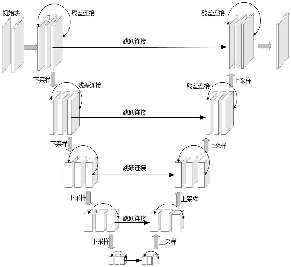
[0036] Hereinafter, the present invention will be further described with reference to the accompanying drawings. This embodiment takes the segmentation of abdominal CT three-dimensional images as an example, and specifically describes the construction process of a segmentation model suitable for abdominal CT three-dimensional images, the model training process, and the image testing process using the model;

[0037] like figure 1 As shown, the specific construction process of the segmentation model described in this embodiment:

[0038] Step S1, data set preprocessing and pretraining, specifically including:

[0039] Step S1.1, according to the segmentation task of the three-dimensional medical image, information encoding is performed as the task encoding information, and the specific operation is: performing information encoding on tasks of different categories. This embodiment takes the abdominal CT three-dimensional image segmentation task as an example. Therefore, there ...


PUM


Abstract

The invention discloses a three-dimensional medical image segmentation model and a training method and application thereof, the model is based on a deep learning convolutional neural network, and the network model is composed of a Prototype calculation module, an encoder decoder module, a task controller module, a dynamic convolution kernel generator module and a dynamic segmentation head module. According to the model, a task controller is designed and a Prototype feature vector is introduced to guide the network to carry out segmentation task discrimination, and different segmentation tasks are flexibly processed by using dynamic convolution, so that the problem of partial marking in the field of medical image segmentation is solved, and the aim of segmenting organs and tumors of multi-category three-dimensional medical images is fulfilled; the problems of excessive parameters and inflexible task expansion in the current mainstream method are solved, and meanwhile, the segmentation result is improved to a certain extent.

Description

technical field [0001] The invention relates to a three-dimensional medical image segmentation model, a training method and application thereof, and belongs to the technical field of image segmentation. Background technique [0002] Automatic segmentation of organs and tumors using computed tomography (CT) is one of the most fundamental but also the most challenging tasks in medical image analysis, and it plays a pivotal role in various computer-aided diagnosis tasks, such as analyzing lesions tasks such as contouring, developing surgical plans, and performing 3D reconstructions. However, due to the limitation of labor cost and expertise, it is difficult to perform voxel-level annotation of multiple organs and tumors in large datasets, so most of the current basic datasets are only used for one type of organ or tumor For the segmentation task, all task-irrelevant organs and tumors are annotated as background. For example, the LiTS dataset only has annotations for liver and...

Claims


Application Information

Patent Timeline
IPC IPC(8): G06T7/10G06T7/11G06T17/00G06N3/04
CPCG06T7/10G06T17/00G06T7/11G06T2207/30096G06T2207/20084G06T2207/20081G06T2207/10081G06T2207/20132G06T2207/10012G06T2207/30084G06N3/045
Inventor 钱誉朱虎邓丽珍
Owner NANJING UNIV OF POSTS & TELECOMM
Why Eureka
Try Eureka