Construction method of chronic hepatitis B cirrhosis prediction model and prediction method of chronic hepatitis B cirrhosis prediction model
A predictive model and technology for chronic hepatitis B, applied in computing models, medical simulation, 2D image generation, etc., can solve the problems of cost, inaccurate prediction, and inability to be widely used, so as to achieve multiple clinical benefits and improve diagnosis performance, effect of improved reproducibility
- Summary
- Abstract
- Description
- Claims
- Application Information
AI Technical Summary
Problems solved by technology
Method used
Image
Examples
specific Embodiment approach 1
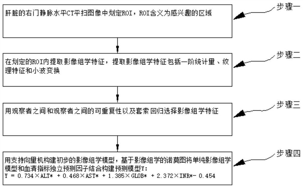
[0057] The construction method of a kind of chronic hepatitis B liver cirrhosis prediction model of the present embodiment, such as figure 1 and Figure 5 As shown, the method is realized through the following steps:
[0058] Step 1. Delineate the ROI in the CT plain scan image at the level of the right portal vein of the liver, wherein the Chinese meaning of ROI is the region of interest, and the full English name is region on interest;
[0059] Step 2, extracting radiomics features in the defined ROI, extracting radiomics features including first-order statistics, texture features and wavelet transform;
[0060] Step 3. Select radiomics features using interobserver and interobserver reproducibility and lasso regression;
[0061] Step 4: Construct a preliminary radiomics model with a support vector machine, and build a prediction model Y based on a nomogram of radiomics by combining a simple radiomics model with independent predictors of serum indicators:
[0062] Y=0.734×...
specific Embodiment approach 2
[0069] The difference from Embodiment 1 is that in the method for constructing a predictive model of chronic hepatitis B cirrhosis in this embodiment, in the step of delineating the ROI in the CT plain scan image at the level of the right portal vein of the liver described in Step 1,
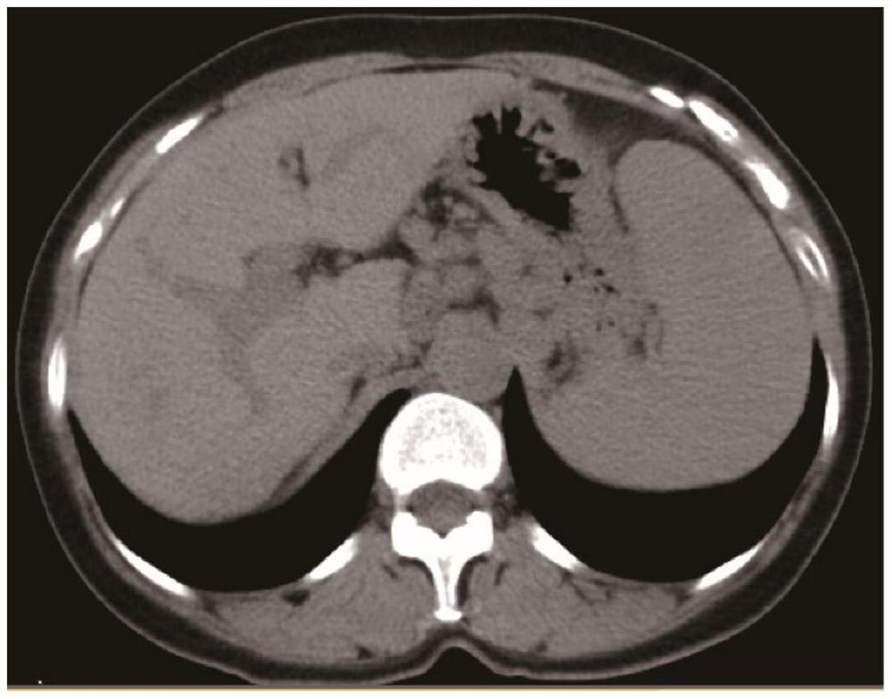
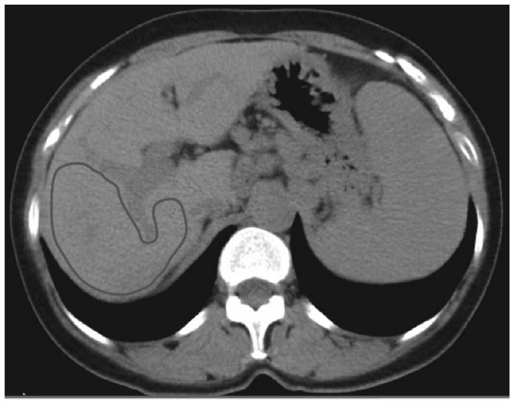
[0070] 3Dslicer software (version 4.8.0; http: / / www.slicer.org) was used to select the ROI area in the right portal vein level CT plain scan images of multiple livers, and the content shown in the ROI area was removed from the plain CT images. The great vessels of the liver are delineated along the margin of the right lobe at the level of the right portal vein; Figure 2a Shown is a plain CT scan image of the liver at the level of the right portal vein, Figure 2b Shown is the delineation of the ROI area in the plain CT image at the level of the right portal vein of the liver; where the area of the ROI ranges from 19 to 106 cm 2 .
specific Embodiment approach 3
[0071] The difference from the second specific embodiment is that in the construction method of a chronic hepatitis B cirrhosis prediction model in this embodiment, the average area of the ROI is 47±15cm 2 .
PUM
 Abstract
Description
Claims
Application Information
 - R&D
- Intellectual Property
- Life Sciences
- Materials
- Tech Scout
- Unparalleled Data Quality
- Higher Quality Content
- 60% Fewer Hallucinations
Browse by: Latest US Patents, China's latest patents, Technical Efficacy Thesaurus, Application Domain, Technology Topic, Popular Technical Reports.
© 2025 PatSnap. All rights reserved.Legal|Privacy policy|Modern Slavery Act Transparency Statement|Sitemap|About US| Contact US: help@patsnap.com


