
Amniotic membrane fixing ring for ophthalmology department

A technology for fixing rings and amniotic membranes, applied in ophthalmic surgery, etc., can solve the problems of poor use effect, strong foreign body sensation in patients, curled amniotic membrane, etc., and achieve the effect of avoiding curling, reducing foreign body sensation, and firm fixation

Inactive Publication Date: 2022-04-29
南京医科大学眼科医院
View PDF0 Cites 1 Cited by
  • Summary
  • Abstract
  • Description
  • Claims
  • Application Information

AI Technical Summary

Problems solved by technology

[0004] 1) The existing amniotic membrane fixation ring fixes the amniotic membrane in a single way, and the amniotic membrane will curl during the operation, making the operation unable to continue;
[0005] 2) It is troublesome to install the existing amniotic membrane fixing ring on the patient's eyes, and the patient's foreign body sensation is strong, and the use effect is poor

Method used


Image

  • Amniotic membrane fixing ring for ophthalmology department
  • Amniotic membrane fixing ring for ophthalmology department
  • Amniotic membrane fixing ring for ophthalmology department

Examples


Embodiment 1

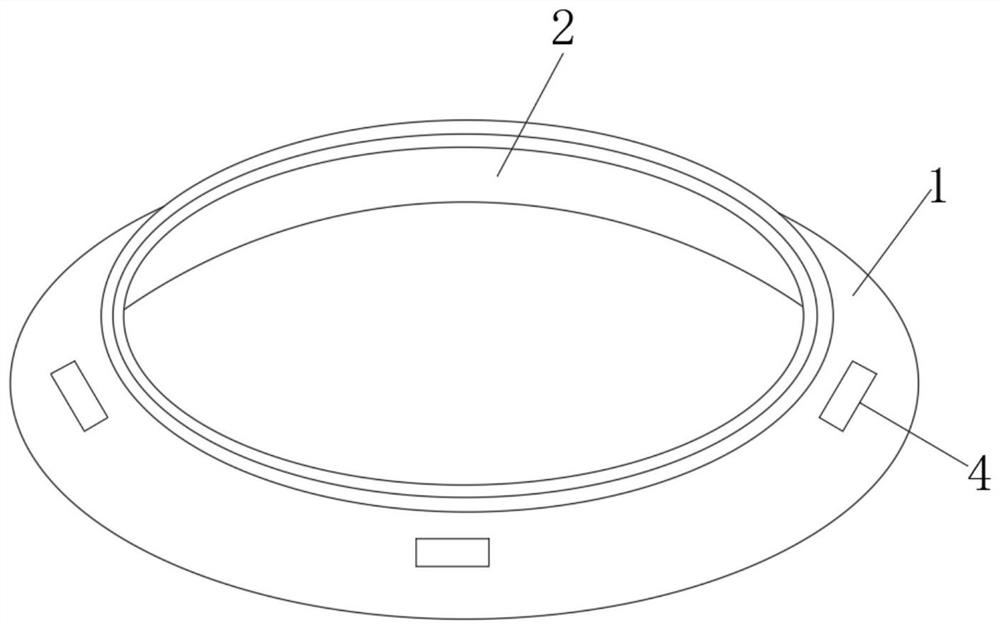
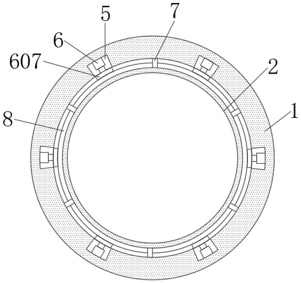
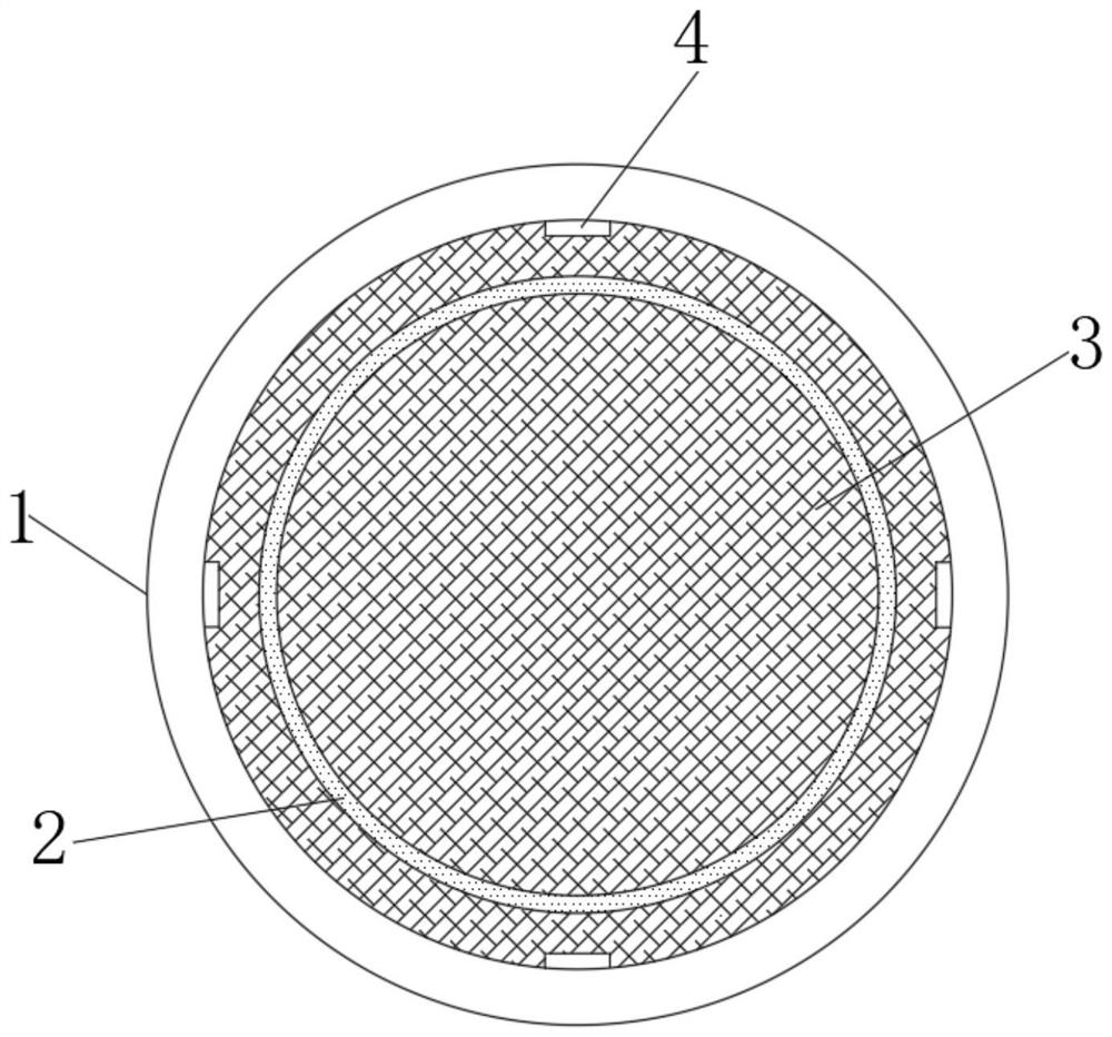
[0028] see Figure 1~5 , an amnion fixation ring for ophthalmology, comprising

[0029] The outer ring piece 1 has four fixing components 4 evenly arranged on the inner side of its outer surface;

[0030] The inner ring piece 2 is installed inside the outer ring piece 1 through the connecting block 7, and a clamping groove 8 is provided between the outer ring piece 1 and the inner ring piece 2;

[0031] The clamping mechanism 6 is fixedly installed in the installation groove 5 provided inside the outer ring piece 1, and the clamping side is placed in the clamping groove 8, and the amniotic membrane 3 is clamped in the clamping groove 8. A clamping mechanism 6 is provided to facilitate fixing the amniotic membrane.

[0032] In the embodiment of the present invention, both the outer ring piece 1 and the inner ring piece 2 are set in a transparent package, so that the medical staff can see clearly the specific situation of the pressing position of the amniotic membrane 3 .

[...

Embodiment 2

[0036] see Figure 1~5 , an amnion fixation ring for ophthalmology, comprising

[0037] The outer ring piece 1 has four fixing components 4 evenly arranged on the inner side of its outer surface;

[0038] The inner ring piece 2 is installed inside the outer ring piece 1 through the connecting block 7, and a clamping groove 8 is provided between the outer ring piece 1 and the inner ring piece 2;

[0039] The clamping mechanism 6 is fixedly installed in the installation groove 5 provided inside the outer ring piece 1, and the clamping side is placed in the clamping groove 8, and the amniotic membrane 3 is clamped in the clamping groove 8. A clamping mechanism 6 is provided to facilitate fixing the amniotic membrane.

[0040] In the embodiment of the present invention, the fixing assembly 4 is composed of an L-shaped fixing block 401 fixed on the surface of the outer ring and a pressing plate 404. The vertical side wall of the L-shaped fixing block 401 is provided with a slide ...


PUM


Abstract

The invention discloses an amniotic membrane fixing ring for ophthalmology. The amniotic membrane fixing ring comprises an outer ring piece, an inner ring piece and a clamping mechanism. The fixing ring has the advantages that the outer edge of the outer ring piece has a certain radian, the radian is matched with the radian of the canthus, the fixing ring can be conveniently installed in the eye, the elastic soft rubber mat is arranged on the lower side, making contact with the eye, of the outer ring piece, and the foreign body sensation of the eye of a patient is reduced; when the amniotic membrane is fixed, the amniotic membrane penetrates into a clamping groove from the bottom side of an inner ring piece, then an arc-shaped clamping block is driven to be tightly attached to the amniotic membrane through the elastic force effect of a second spring, and therefore the amniotic membrane is clamped and fixed, and the excessive amniotic membrane penetrates out of the clamping groove and extends into a fixing assembly arranged on the outer surface of an outer ring piece; a pressing plate is extruded through a first spring, so that the pressing plate tightly presses the amnion extending out of the inner ring piece, the amnion is conveniently limited, the amnion is firmly fixed in a double-fixing mode, and the situation that the amnion is curled in the operation process is avoided.

Description

technical field [0001] The invention relates to an amniotic membrane fixing ring, in particular to an amniotic membrane fixing ring for ophthalmology, and belongs to the technical field of ophthalmic treatment auxiliary equipment. Background technique [0002] The amniotic membrane is a translucent membrane located in the innermost layer of the placenta. It is smooth, free of blood vessels, nerves and lymphatic tissues, and has certain elasticity. In conventional amniotic covering surgery, under local infiltration anesthesia or general anesthesia, the amniotic membrane is fixed to the conjunctiva or superficial scleral tissue with intermittent or continuous 10-0 nylon sutures. Each operation takes about 40-60 minutes, and the sutures The thread is easy to cause postoperative conjunctival granuloma. At the same time, because the amniotic membrane is thin, with a thickness of about 0.02-0.05mm, the amniotic membrane is often pulled by forceps or needles during the operation, ...

Claims


Application Information

Patent Timeline
Patent Type & Authority Applications(China)
IPC IPC(8): A61F9/007
CPCA61F9/007
Inventor 杨卫华李柯然蒋沁曹国凡王成虎纪玉珂
Owner 南京医科大学眼科医院
Learn More