
Mesh bone cement spacer for wrapping cancellous bone grafting for bone defect and preparation method of mesh bone cement spacer

A bone cement and cancellous bone technology, applied in bone implants, manufacturing tools, additive manufacturing, etc., can solve the problems of simultaneous bone grafting and no space for bone grafting

Active Publication Date: 2022-04-19
WUXI NO 9 PEOPLES HOSPITAL
View PDF10 Cites 0 Cited by
  • Summary
  • Abstract
  • Description
  • Claims
  • Application Information

AI Technical Summary

Problems solved by technology

The present invention can overcome the defects of filling the solid bone cement interval with traditional induction membrane technology, there is no space for bone grafting, no bone grafting at the same time, and the two-stage operation that needs to take out the bone cement interval and re-implant bone, and realize simultaneous filling of the bone cement interval and wrapping The purpose of the modified induced membrane technique of bone grafting in one-stage surgery to repair bone defects

Method used


Image

  • Mesh bone cement spacer for wrapping cancellous bone grafting for bone defect and preparation method of mesh bone cement spacer
  • Mesh bone cement spacer for wrapping cancellous bone grafting for bone defect and preparation method of mesh bone cement spacer
  • Mesh bone cement spacer for wrapping cancellous bone grafting for bone defect and preparation method of mesh bone cement spacer

Examples


Embodiment 1

[0034] like Figure 1-Figure 4 shown:

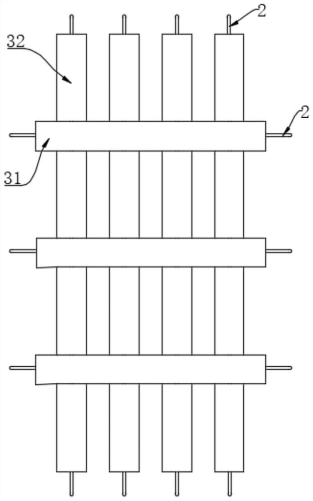
[0035] After debridement, the bone defect in the upper segment of the human tibia has a length of 100mm and a diameter of 60mm, and the two ends of the bone defect are fixed with steel plates 4; the mesh wrapping the bone defect needs to wrap the two stumps by 1cm respectively, so the width of the mesh is 100+10+10=120mm , the length is 60×3.1415=188mm.

[0036] A method for preparing a mesh bone cement spacer wrapped with cancellous bone graft for bone defect, comprising the following steps:

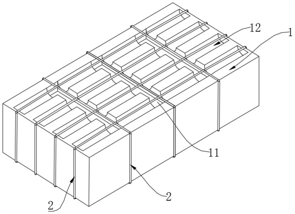
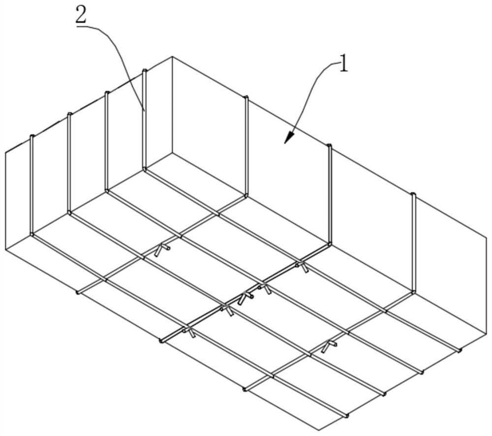
[0037]Step 1: Use foam material to make a mold 1 with 4 horizontal grooves and 30 vertical grooves through 3D printing technology; wherein, the length of each horizontal groove is 188mm, the width is 1mm, and the depth is 1mm; each vertical groove The length of the groove is 120mm, the width is 2mm, and the depth is 2mm; the distance between every two adjacent transverse grooves is 25mm; the distance between every two adjacent longitudinal groov...

Embodiment 2

[0048] like Figure 1-Figure 4 Shown:

[0049] The diameter of the mid-section of the femur of SD rats is 4mm, and the bone defect of the mid-section of the femur of SD rats is made with a bone defect length of 10mm, that is, a 4mm×10mm bone defect, and the two ends of the bone defect are fixed with steel plates 4; the mesh wrapping the bone defect needs to be wrapped separately The two residual sections are 2mm, so the length of the net is 10+2+2=14mm, and the width is 4×3.141=12mm.

[0050] A method for preparing a mesh bone cement spacer wrapped with cancellous bone graft for bone defect, comprising the following steps:

[0051] Step 1: choose a cardboard material to press mold and make a mold 1 with 3 transverse grooves and 5 longitudinal grooves; wherein, the length of each transverse groove is 14mm, the width is 1mm, and the depth is 0.8mm; The length of each longitudinal groove is 12mm, the width is 1.5mm, and the depth is 0.8mm; the distance between every two adjacen...

Embodiment 3

[0061] like Figure 1-4 Shown:

[0062] After debridement, the length of the bone defect in the middle part of the human femur is 60mm, and the diameter of the femur is 50mm. The two ends of the bone defect are fixed with steel plates 4; =80mm, the length is 50×3.14=160mm.

[0063] A method for preparing a mesh bone cement spacer wrapped with cancellous bone graft for bone defect, comprising the following steps:

[0064] Step 1: Use resin material to make a mold 1 with 3 horizontal grooves and 32 vertical grooves through 3D printing technology; wherein, the length of each horizontal groove is 160mm, the width is 1mm, and the depth is 1mm; each vertical groove The length of the groove is 80mm, the width is 3mm, and the depth is 2mm; the distance between every two adjacent transverse grooves is 20mm; the distance between every two adjacent longitudinal grooves is 2mm;

[0065] Step 2: Use a sewing needle to pass No. 1 medical non-absorbable suture through the center of the le...


PUM

PropertyMeasurementUnit
lengthaaaaaaaaaa
widthaaaaaaaaaa
depthaaaaaaaaaa

Abstract

The invention provides a preparation method of a reticular bone cement spacer for wrapping cancellous bone grafting for bone defect, which comprises the following steps of: 1, manufacturing a mold with transverse and longitudinal grooves, and presetting ribs for controlling mesh gaps in the grooves; and 2, filling the transverse and longitudinal grooves with bone cement, and after the bone cement is shaped and hardened, taking out the reticular bone cement intervals with the ribs. The net-shaped bone cement prepared by the preparation method disclosed by the invention is spaced, and relatively thin transverse connection can be broken, so that the net has moldability and can be used for wrapping bone defects with different shapes and sizes; due to the fact that ribs formed by sutures or thin steel wires are arranged in the transverse intervals and the longitudinal intervals, the intervals cannot be scattered after being broken, the hole gap size can be constantly kept, and the excellent bone grafting material wrapping effect is achieved; the reticular bone cement spacer can stimulate surrounding soft tissue to form an induction membrane, the biological osteogenesis promoting effect is achieved, the bone healing effect is better compared with a pure mechanical wrapping bone grafting method, and the good repeatability and the satisfactory osteogenesis effect are achieved.

Description

technical field [0001] The invention relates to the technical field of biomedical materials, in particular to a mesh bone cement spacer for wrapping cancellous bone graft for bone defect and a preparation method thereof. Background technique [0002] Segmental bone defects caused by resection of high-energy trauma, bone infection, bone tumors, and congenital skeletal deformities are common in clinical practice, and the repair of large bone defects is still challenging. Traditional free bone grafting is only suitable for small-segment bone defects, and is considered unsuitable for large-segment bone defects, because the bone graft material is prone to loosening and bone resorption after traditional free bone grafting in large-segment bone defects, and the nonunion rate is high. Recently, improved free bone grafting methods have emerged, such as wrapped bone grafting techniques such as titanium mesh, wire mesh, and fascia that wrap bone graft materials such as cancellous bone....

Claims


Application Information

Patent Timeline
Patent Type & Authority Applications(China)
IPC IPC(8): A61F2/28A61F2/30
CPCA61F2/28A61F2/30724A61F2/30907A61F2/30942A61F2002/2835A61F2002/30909A61F2002/3093A61F2002/30985A61F2002/30772A61F2002/307
Inventor 马运宏卜凡玉殷渠东
Owner WUXI NO 9 PEOPLES HOSPITAL
Learn More