
Method and system for detecting breast cancer area of pathological image based on DenseNet network

A technology for pathological images and breast cancer, applied in mammography, neural learning methods, biological neural network models, etc., can solve time-consuming problems and achieve the effect of suppressing isolated noise

Active Publication Date: 2022-02-11
XINXIANG MEDICAL UNIV
View PDF6 Cites 1 Cited by
  • Summary
  • Abstract
  • Description
  • Claims
  • Application Information

AI Technical Summary

Problems solved by technology

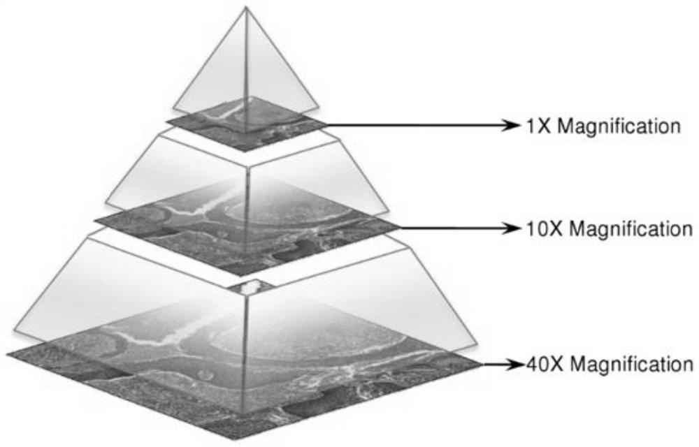
Pathology images are very large gigapixel images with an image size of 10 6 x10 6 , which is a very time-consuming and challenging task for pathologists

Method used


Image

  • Method and system for detecting breast cancer area of pathological image based on DenseNet network
  • Method and system for detecting breast cancer area of pathological image based on DenseNet network
  • Method and system for detecting breast cancer area of pathological image based on DenseNet network

Examples


Embodiment Construction

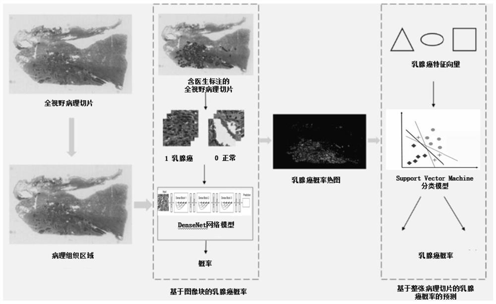
[0064] This example presents a breast cancer area detection method based on a DenseNet network-based full-field breast cancer sentinel lymph node pathological image. The DenseNet network model is used to learn the characteristics of breast cancer pathological image blocks, generate a full-field breast cancer probability heat map, and calculate breast cancer. Cancer feature vector, using SVM to predict the probability of occurrence of breast cancer regions, to achieve automatic detection of breast cancer regions.

[0065] The hardware environment of this embodiment is: Intel Xeon E5-2678v3 dual processors, memory 32.0GB, graphics card RTX2080Ti 2 pieces, software environment Ubuntu 16.04, Python 3.5, Tensorflow, OpenSlide, SciKit, NumPy.

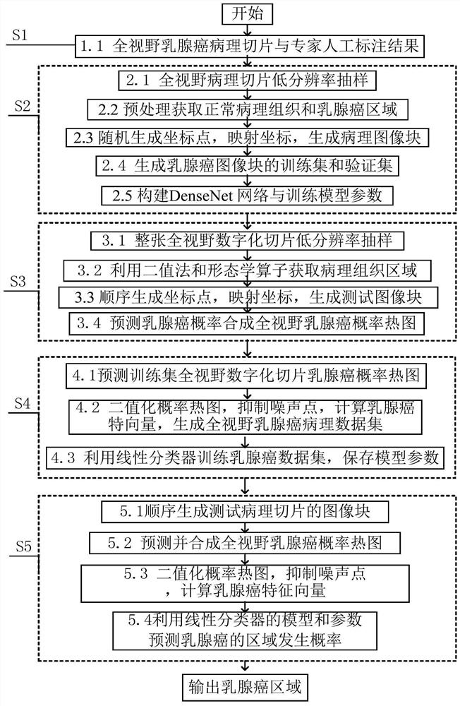
[0066] This embodiment presents a breast cancer region detection method based on a DenseNet network-based full-field breast cancer sentinel lymph node pathological image, and its flow chart is as follows figure 1 As shown, follow the steps b...


PUM


Abstract

The invention relates to a breast cancer area detection method and system for a full-view breast cancer sentinel node pathological image based on a DenseNet network, and the method comprises the steps: 1) obtaining the full-view breast cancer sentinel node pathological image, and manually marking a breast cancer metastasis area; 2) automatically generating a training set and a verification set by using the full-view breast cancer sentinel lymph node pathological image and the labeling result, and training by using a DenseNet network model; 3) predicting a single full-view breast cancer pathology image to obtain a breast cancer probability heat map; 4) calculating breast cancer probability heat maps of the full-view breast cancer pathological images of all the training sets, performing binaryzation and isolated noise suppression, calculating breast cancer feature vectors, and performing training by using a linear classifier; 5) calculating a breast cancer probability heat map for the full-view pathological image of the test set, carrying out binaryzation and isolated noise suppression, calculating a breast cancer feature vector, and predicting the occurrence probability of the breast cancer region, thereby realizing automatic detection of the breast cancer region.

Description

technical field [0001] The invention belongs to the technical field of pathological image processing, and specifically relates to a breast cancer region detection method and system based on a DenseNet network-based full-field sentinel lymph node pathological image of breast cancer. The method can automatically analyze the full-field sentinel lymph node pathological image of breast cancer, Automated detection of breast cancer regions. Background technique [0002] Breast cancer is the one with the highest incidence rate among all malignant tumors in women, accounting for about 17% of all malignant tumors in China, seriously endangering women's health and even life-threatening. The cause of breast cancer is not yet fully understood, and the incidence rate is high, but if detected early, the possibility of cure is very high. Early detection and diagnosis of breast cancer is the key to improving the curative effect. In the process of diagnosis and treatment of breast cancer, m...

Claims


Application Information

Patent Timeline
IPC IPC(8): A61B5/00G06V10/25G06V10/22G06V10/42G06K9/62G06N3/04G06N3/08
CPCA61B5/7264A61B5/7267A61B5/7203A61B5/4312G06N3/08G06N3/045G06F18/2414G06F18/2411G06F18/2415Y02A90/10
Inventor 王昌张文超闫岑张业宏申杰奋秦鑫赵俊强于毅吴阳王辰
Owner XINXIANG MEDICAL UNIV
Learn More