Eye fundus hydrops segmentation method of OCT image

An image and effusion technology, applied in the field of fundus effusion segmentation of OCT images, can solve the problems of retinal layer damage, retinal layer topology and shape influence, retinal layer reflectivity changes, etc., to achieve the effect of improving accuracy

Pending Publication Date: 2021-12-03
JILIN UNIV
View PDF0 Cites 0 Cited by
  • Summary
  • Abstract
  • Description
  • Claims
  • Application Information

AI Technical Summary

Problems solved by technology

[0006] The presence of fundus fluid can lead to extensive damage to the retinal layers. First, the topology and morphology of the retinal layers

Method used


Image

  • Eye fundus hydrops segmentation method of OCT image
  • Eye fundus hydrops segmentation method of OCT image
  • Eye fundus hydrops segmentation method of OCT image

Examples


Embodiment Construction

[0100] In order to make the object, technical solution and advantages of the present invention clearer, the present invention will be further described in detail below in conjunction with the accompanying drawings and embodiments. It should be understood that the specific embodiments described here are only used to explain the present invention, not to limit the present invention.

[0101] The specific implementation of the present invention will be described in detail below in conjunction with specific embodiments.

[0102] 1. Fundus fluid segmentation step, the OCT image fundus fluid segmentation step includes:

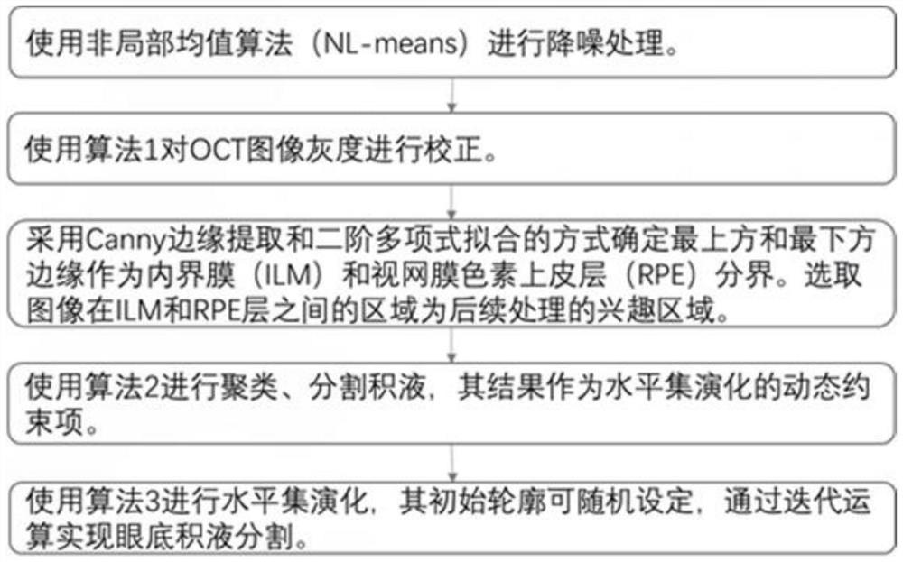
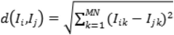
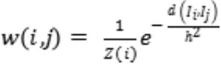
[0103] 1) Use non-local mean algorithm (NL-means) for noise reduction processing;

[0104] 2) Use Algorithm 1 to correct the gray scale of the OCT image;

[0105] 3) Using Canny edge extraction and second-order polynomial fitting to determine the uppermost and lowermost edges as the boundary between the inner limiting membrane (ILM) and the retinal pigment epithel...


PUM


Abstract

The invention is suitable for the technical field of medical image processing, and provides a fundus hydrops segmentation method of an OCT image, which comprises the following steps of: 1, carrying out noise reduction processing on the input OCT image by using a non-local mean algorithm; 2, correcting the gray level of the OCT image by using an algorithm 1; 3, adopting a Canny edge extraction and second-order polynomial fitting mode to determine the uppermost edge and the lowermost edge as the boundary of the inner boundary membrane and the retinal pigment epithelium, and selecting the region, between the inner boundary membrane and the retinal pigment epithelium, of the image as the region of interest of subsequent processing; 4, clustering by using an improved fuzzy clustering algorithm, and segmenting effusion; and 5, performing level set evolution by using a level set algorithm, and realizing fundus effusion segmentation through iteration. According to the method, pixel data needing to be processed is reduced under the condition that the image quality is guaranteed, the fuzzy clustering result serves as a dynamic constraint item of the level set method to control evolution of the level set, the method is not affected by a randomly-set initial level set, and the accuracy is effectively improved.

Description

technical field [0001] The invention belongs to the technical field of medical image processing, in particular to a fundus fluid segmentation method of an OCT image. Background technique [0002] The retina is a layered structure, and each layer of tissue in the eyeball has a certain thickness. The image of the layered structure of the retina captured by optical equipment can be used to distinguish between healthy and unhealthy retinas, and further analyze the characteristics of the diseased retina. Retinal images reflect the patient's condition and provide effective diagnostic measures. [0003] Optical coherence tomography (OCT) imaging technology uses near-infrared light to realize the collection of retinal layered images. Scanning the retina with a laser takes and produces an image that shows the layers of the retina in great detail. The use of OCT imaging technology can detect eye disease abnormalities, diagnose retinal diseases and prevent irreparable ophthalmic dama...

Claims


Application Information

Patent Timeline
IPC IPC(8): G06T7/11G06T7/13G06T5/00G06T7/90G06K9/62
CPCG06T7/11G06T7/13G06T5/002G06T7/90G06T2207/10101G06T2207/30041G06F18/23
Inventor 郭晓新张宝亮王韩
Owner JILIN UNIV
Why Eureka
Try Eureka