Method for detecting jugular vein ball socket bone wall deficiency in temporal bone CT image

A jugular ball and bone wall technology, which is applied in the field of abnormal detection of jugular ball socket bone wall in temporal bone CT, can solve the problems of difficult classification, target detection, and feature extraction, etc., and achieve low requirements for the number of abnormal data , high promotional effect

Pending Publication Date: 2021-11-12
BEIJING UNIV OF TECH
View PDF0 Cites 0 Cited by
  • Summary
  • Abstract
  • Description
  • Claims
  • Application Information

AI Technical Summary

Problems solved by technology

For the abnormal detection of jugular vein ball-socket bone wall loss, there may be the following problems: (1) The jugular vein ball-socket target size is small in temporal bone CT, and the bone wall loss lesion target is even smaller, which makes target detection difficult and a large number of false positive samples are likely to be generated; ( 2) Abnormal samples are far less than normal samples, the categories are extremely unbalanced, features are difficult to extract, and categories are difficult to distinguish
Anomaly detection based on self-encoder has strong network generalization ability. When an abnormal image is input, the decoded image is very close to the input image, and it is difficult to distinguish between normal and abnormal.
Codec networks are usually used in unsupervised tasks without the guidance of abnormal data

Method used


Image

  • Method for detecting jugular vein ball socket bone wall deficiency in temporal bone CT image
  • Method for detecting jugular vein ball socket bone wall deficiency in temporal bone CT image
  • Method for detecting jugular vein ball socket bone wall deficiency in temporal bone CT image

Examples


Embodiment Construction

[0022] Exemplary implementation examples of the present invention will be described in more detail below with reference to the accompanying drawings. Although exemplary embodiments of the present invention are shown in the drawings, it should be understood that the invention may be embodied in various forms and should not be limited to the embodiments set forth herein. Rather, these embodiments are provided for more thorough understanding of the present invention and to fully convey the scope of the present invention to those skilled in the art.

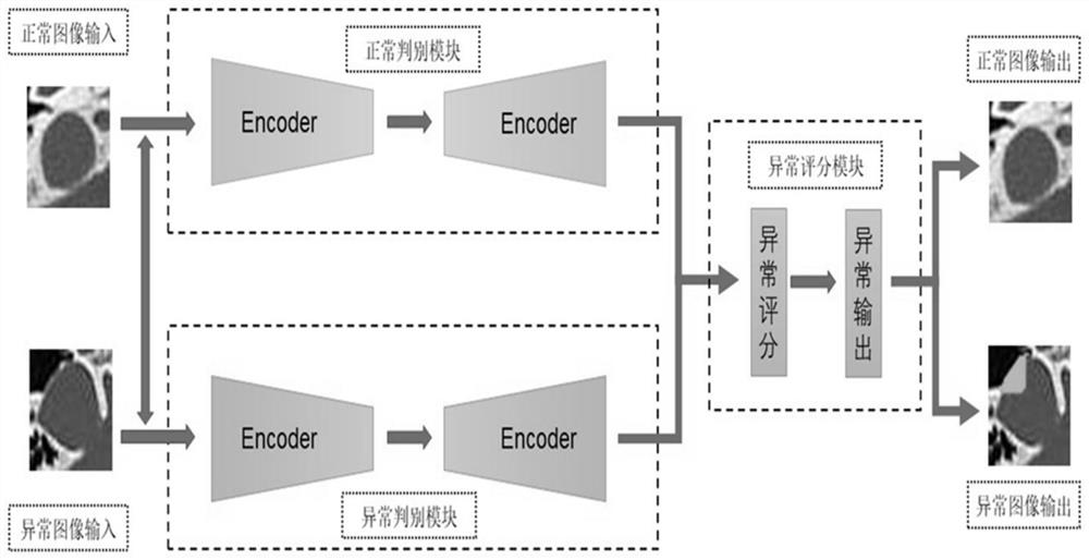
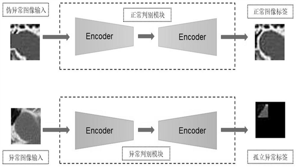
[0023] This method makes full use of a small amount of rare anomaly data to mine the fundamental and unique characteristics of anomalies. In the few but clear abnormal samples, fully highlight the characteristics of abnormal parts, reduce the annihilation of abnormal features in the feature extraction process, so that the model can have a higher sensitivity to abnormal parts, so that the network is more sensitive to normal features a...


PUM


Abstract

The invention provides a method for detecting jugular vein ball socket bone wall deletion abnormity in a temporal bone image. The method can be used for computer-aided pulsatile tinnitus diagnosis. According to the method, a small amount of rare abnormal data is fully utilized, abnormal prior information is introduced into a network model, abnormal features are highlighted, a difference between the abnormal features is increased, and the overall detection accuracy is improved. Through a targeted data enhancement mode and an innovative data use means, a reasonable proxy task is designed, so that the network model gives full play to a advantage of being good at prediction, and detection accuracy is improved. Compared with an existing method, the method provided by the invention does not depend on a large amount of labeled data any more, and has advantages of being easier to implement and popularize.

Description

technical field [0001] The invention relates to the field of medical image processing, in particular to a method for detecting the abnormality of bone wall loss of the jugular vein ball socket image in temporal bone CT. Background technique [0002] Tinnitus is manifested as the abnormal perception of sound by the human body without external stimuli. It is a common and frequently-occurring disease in otology. Among them, pulsatile tinnitus (Pulsatile Tinnitus, PT) accounts for about 4%-10% of tinnitus patients. In the past ten years, with economic development and people's emphasis on health, the demand for diagnosis and treatment of patients has surged. With the popularization of temporal bone imaging technology, the imaging method for pulsatile tinnitus has become an important technical means. [0003] Temporal bone CT is the main method for diagnosing osseous lesions in pulsatile tinnitus. Temporal bone CT can display multiple fine structures such as the middle ear and i...

Claims


Application Information

Patent Timeline
Patent Type & Authority Applications(China)
IPC IPC(8): A61B6/03
CPCA61B6/032A61B6/50A61B6/505A61B6/5205A61B6/5211A61B6/5217Y02T10/40
Inventor 李晓光周祎超张辉卓力
Owner BEIJING UNIV OF TECH
Why Eureka
Try Eureka