
Automatic brain infarction detection system on magnetic resonance imaging and operation method thereof

A radiography and brain technology, applied in the field of automatic brain infarction detection system, can solve the problems that MRI examination cannot detect, brain tissue energy metabolism damage, etc.

Pending Publication Date: 2021-10-19
NAT CENT UNIV +2
View PDF0 Cites 0 Cited by
  • Summary
  • Abstract
  • Description
  • Claims
  • Application Information

AI Technical Summary

Problems solved by technology

[0002] After cerebral infarction, the energy metabolism of brain tissue is destroyed, and a large amount of extracellular water enters the cells, causing the increase of intracellular water molecules and the decrease of extracellular water molecules, resulting in restricted diffusion, but the water content in the ischemic area will not Changes are only caused by changes in the water content inside and outside the cells, resulting in limited diffusion, so conventional MRI examinations such as T1, T2, FLAIR... etc. are often unable to detect

Method used


Image

  • Automatic brain infarction detection system on magnetic resonance imaging and operation method thereof
  • Automatic brain infarction detection system on magnetic resonance imaging and operation method thereof

Examples


Embodiment Construction

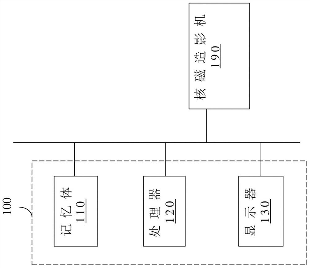
[0028] In order to make the description of the present invention more detailed and complete, reference may be made to the attached drawings and various embodiments described below, and the same numbers in the drawings represent the same or similar elements. On the other hand, well-known elements and steps have not been described in the embodiments in order to avoid unnecessarily limiting the invention.

[0029] In the embodiments and claims, the description involving "connection" can generally refer to an element being indirectly coupled to another element through other elements, or an element is directly connected to another element without passing through other elements .

[0030] In the implementation and claims, the description related to "connection" can generally refer to a component that indirectly communicates with another component through wired and / or wireless communication, or a component does not need to pass through other components And the entity is connected to...


PUM


Abstract

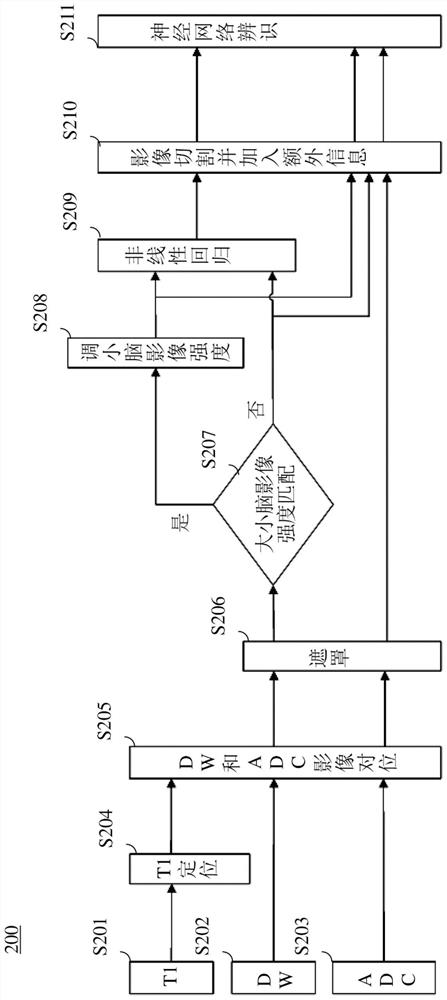
The present disclosure provides an operating method of an automatic brain infarction detection system on magnetic resonance imaging (MRI), which includes steps as follows. Images corresponding to different slices of a brain of a subject are received from the MRI machine. The image mask process is performed on first and second images of the images. It is determined whether the cerebellum image intensity and the brain image intensity in the first image are matched. When the cerebellum image intensity and the brain image intensity are not matched, the cerebellar image intensity in the first image is adjusted. The first image is processed through a nonlinear regression to obtain a third image. A neural network identify an infarct region by using the first, second and third images that are cut. By means of the technical scheme, the position of the focus on magnetic resonance imaging is accurately detected, and diagnosis quantification of a physician on magnetic resonance imaging is more efficiently helped clinically.

Description

technical field [0001] The present invention relates to a system and its operating method, and in particular to a magnetic resonance imaging automatic cerebral infarction detection system and its operating method. Background technique [0002] After cerebral infarction, the energy metabolism of brain tissue is destroyed, and a large amount of extracellular water enters the cells, causing the increase of intracellular water molecules and the decrease of extracellular water molecules, resulting in restricted diffusion, but the water content in the ischemic area will not Changes are only caused by changes in the water content inside and outside the cells, resulting in restricted diffusion, so conventional MRI examinations such as T1, T2, FLAIR... etc. are often unable to detect. [0003] Therefore, how to provide an automatic cerebral infarction detection system and its operation method has become an important issue. Contents of the invention [0004] The present invention p...

Claims


Application Information

Patent Timeline
Patent Type & Authority Applications(China)
IPC IPC(8): G16H50/20G06T7/00G06T5/30G06N3/04G06N3/08
CPCG16H50/20G06T7/0012G06T7/97G06T5/30G06N3/08G06T2207/10088G06T2207/20084G06T2207/20081G06T2207/30016G06T2207/30096G06N3/045G06T7/0016G06T5/009
Inventor 蔡章仁彭徐钧陈右纬蔡孟宗王国伟郭叶璘
Owner NAT CENT UNIV
Learn More