
Fundus image microhemangioma detection device and method thereof and storage medium

A technology for microvascular tumors and fundus images, which is applied in image enhancement, image analysis, image data processing, etc., and can solve the problems of low detection accuracy, low accuracy, and difficult integration and use.

Pending Publication Date: 2021-10-15
UNIV OF ELECTRONIC SCI & TECH OF CHINA
View PDF0 Cites 1 Cited by
  • Summary
  • Abstract
  • Description
  • Claims
  • Application Information

AI Technical Summary

Problems solved by technology

These methods have not analyzed the characteristics of MA itself, so there are still characteristics of low accuracy and weak robustness.
Therefore, the current microvascular tumor detection method based on computer image processing technology still has problems such as low robustness, low detection accuracy, and difficulty in integrated use.

Method used


Image

  • Fundus image microhemangioma detection device and method thereof and storage medium
  • Fundus image microhemangioma detection device and method thereof and storage medium
  • Fundus image microhemangioma detection device and method thereof and storage medium

Examples


Embodiment Construction

[0057] The present invention will be further described in detail below in conjunction with test examples and specific embodiments. But this should not be interpreted as that the scope of the above-mentioned theme of the present invention is limited to the following embodiments, and all technologies realized based on the content of the present invention all belong to the scope of the present invention.

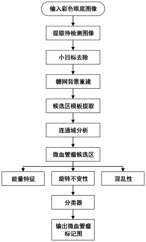
[0058] The present invention proposes a method for detecting microvascular tumors in fundus images, which can detect microvascular tumors in fundus images, and has high specificity and sensitivity. The entire algorithm design process is as follows: figure 1 shown, including steps:

[0059] In the above technical solution, the step 1 specifically has the following steps:

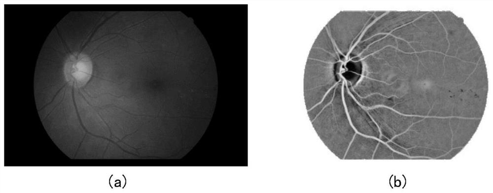
[0060] Step 1.1: Extract the green channel image from the input color fundus image, and reflect it to obtain the image I to be detected. In this example, the size of the input color fundus image is 2544×1696...


PUM


Abstract

The invention discloses a fundus image microhemangioma detection device and a method and a storage medium, and relates to the field of medical image processing and machine vision application. The method comprises the following steps: firstly, a to-be-detected image is extracted from an input color retina image, and then a micro-hemangioma candidate region template and positive and negative sample pictures are obtained through operations such as small target removal and geodesic expansion; energy features are extracted for the micro-hemangioma candidate area, features are manually designed for the micro-hemangioma form, and finally, traditional features and the manually designed features are cascaded; and the features and the labels are sent to a trained classifier for classification, so that the position of the micro-hemangioma is detected from the sugar net image. According to the method, the tiny target microhemangioma can be detected through the diabetes eye fundus image, the accuracy is high, and an eye fundus doctor can be assisted to observe the existence of the microhemangioma more conveniently.

Description

technical field [0001] The invention relates to a device, method and storage medium for detecting microvascular tumors in fundus images. Background technique [0002] Diabetic patients will develop retinal lesions in the late stage. Microaneurysm (MA) in fundus images is the initial symptom of diabetic retinopathy (sugar net). . Manual detection of microvascular tumors on fundus images mainly relies on ophthalmologists to directly observe retinal images. However, due to the complex structure of the retina, the small area of ​​microvascular tumors and low local contrast, it is time-consuming, laborious, and remote to observe microvascular tumors with human eyes. There is a lack of experienced ophthalmologists in the region, and the automatic detection of retinal microvascular tumors through computer vision technology will help relieve the pressure on ophthalmologists, and at the same time contribute to the sinking of medical resources, which has far-reaching medical signific...

Claims


Application Information

Patent Timeline
IPC IPC(8): G06T7/00G06T7/187G06T7/194G06T5/00G06T5/30
CPCG06T7/0012G06T7/187G06T7/194G06T5/30G06T2207/30101G06T5/70Y02A90/10
Inventor 邓佳坤彭真明赵学功程晓斌魏浩然曲超唐普英
Owner UNIV OF ELECTRONIC SCI & TECH OF CHINA
Learn More