A biomarker analysis method based on multiple immunohistochemical techniques and its application

A biomarker and immunohistochemical technology, applied in the field of biological detection and analysis, can solve problems such as the inability to explain the spatial relationship of tumor cells well, and achieve the effect of reducing the complicated work

Active Publication Date: 2022-04-08
深圳裕泰抗原科技有限公司
View PDF7 Cites 0 Cited by
  • Summary
  • Abstract
  • Description
  • Claims
  • Application Information

AI Technical Summary

Problems solved by technology

[0006] However, current studies on immuno-oncology focus on the role of specific immune cell subsets in their spatial environment, for example, through multiplex imaging, mass cytometry, and single-cell RNA sequencing, but they are not fully developed. Using information such as spatial coordinate data, the spatial relationship between tumor cells and surrounding elements cannot be well explained

Method used


Image

  • A biomarker analysis method based on multiple immunohistochemical techniques and its application
  • A biomarker analysis method based on multiple immunohistochemical techniques and its application
  • A biomarker analysis method based on multiple immunohistochemical techniques and its application

Examples


Embodiment 1

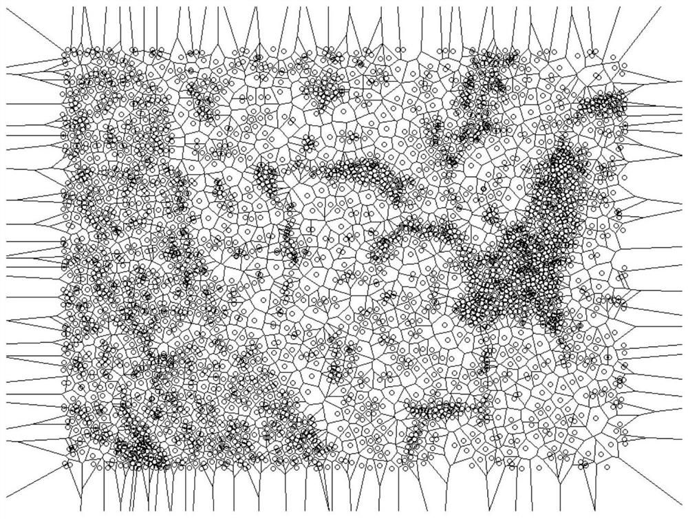
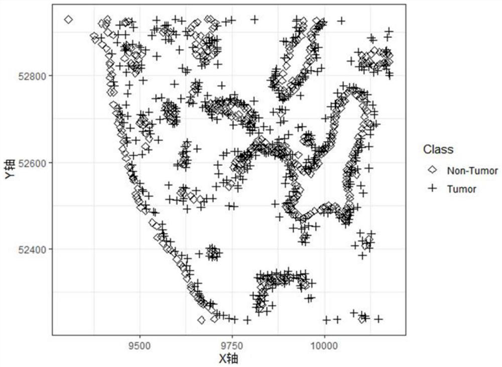
[0075] The sample used in this example is a tumor tissue sample from a clinical lung cancer patient (Patient1) tested in our laboratory. Multiplex immunohistochemical staining (e.g., 7-color MOTiF for lung cancer) TM PD-1 / PD-L1 Panel kit, including 6 kinds of antibodies targeting FoxP3, PD-L1, PanCK, PD-1, CD8, CD68), used to label fluorescent positive cells, and processed by VECTRA POLARIS system Scan to obtain immunohistochemical microscopic panorama. Select the target area (area including tumor inner cells, tumor border cells, and stromal cells) through Phenochart, import it into the inForm system, use the official predefined rule-based lung cancer algorithm to obtain histomorphological classification and cell phenotype, and export the data for biological use. Marker analysis and calculation.

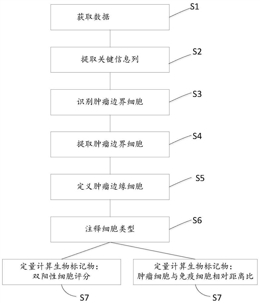
[0076] The biomarker analysis steps are as follows:

[0077] 1. Read data: import "*cell_seg_data.txt" data to Rstudio;

[0078] 2. Extract data: extract key information columns ...

Embodiment 2

[0089] The sample used in this example is the tumor tissue sample of another clinical lung cancer patient (Patient2) tested in our laboratory. Multiple immunohistochemical staining (7 colors and 6 labels: FoxP3, PD-L1, PanCK, PD-1, CD8, CD68) of the patient's tumor tissue sample has been completed, and the immunohistochemical staining was obtained by whole-slice scanning with the VECTRA POLARIS system Microscopic panorama. Select the target area (area including tumor inner cells, tumor border cells, and stromal cells) through Phenochart, import it into the inForm system, use the official predefined rule-based lung cancer algorithm for histomorphological classification and cell typing, and export data for biological use Marker analysis and calculation.

[0090] The biomarker analysis steps are as follows:

[0091] 1. Read data: import "*cell_seg_data.txt" data to Rstudio;

[0092]2. Extract data: extract key information columns (including: histomorphological classification c...


PUM


Abstract

The invention provides a biomarker analysis method based on multiple immunohistochemical techniques and its application. Among them, the biomarker analysis method uses the histological morphology, cell phenotype and corresponding spatial coordinate information of tumor tissue samples in the image recognition system, and compares the two biomarkers through the double positive cell score and the relative distance ratio of tumor-immune suppression. Automated analysis of the tumor immune microenvironment. The present invention also provides devices and devices for implementing the method, as well as corresponding computer-readable media. The invention can more accurately and efficiently characterize the spatial connection state of the tumor immune microenvironment, so that the relevant indicators of the tumor immune microenvironment can more effectively evaluate the curative effect of immunotherapy, and meet the requirements of multiple immunohistochemical techniques in clinical pathological work and scientific research. The need for tumor immune microenvironment analysis.

Description

technical field [0001] The invention relates to the field of biological detection and analysis, in particular to a biomarker analysis method based on multiple immunohistochemical techniques and its application. Background technique [0002] Immunohistochemistry (Immunohistochemistry), also known as immunohistochemistry, is a tissue and cell in situ detection technique for the distribution of antigens (or antibodies) in tissues using labeled specific antibodies (or antigens). As a mature, reliable, economical and widely used technical means, it is currently widely used in clinical pathological diagnosis and scientific research, for the judgment of tumor tissue source, assessment of tumor risk, selection of treatment methods, and various experiments. It plays an irreplaceable role in observing the level and distribution range of antibody (or antigen) expression in tissues and cells. Immunohistochemistry usually includes traditional immunohistochemistry as well as multiplex im...

Claims


Application Information

Patent Timeline
Patent Type & Authority Patents(China)
IPC IPC(8): G16B40/00
CPCG16B40/00
Inventor 李晓婷金皓玄王佳茜苏小凡
Owner 深圳裕泰抗原科技有限公司
Why Eureka
Try Eureka