
Gastrointestinal endoscope polyp removal auxiliary collector and method

A collector and polyp technology, applied in the direction of endoscopes, heated surgical instruments, surgical instrument parts, etc., can solve the problems of polyp structure damage, cumbersome and inconvenient operation, and affect polyp pathological detection data, so as to improve accuracy, The effect of protecting the quality structure and data accuracy

Active Publication Date: 2021-09-07
HUZHOU MATERNITY & CHILD CARE HOSPITAL
View PDF25 Cites 3 Cited by
  • Summary
  • Abstract
  • Description
  • Claims
  • Application Information

AI Technical Summary

Problems solved by technology

Existing excision and collection of polyps, among them, high-frequency electrocoagulation is the most widely used method. In the process of excision of pedicled polyps or narrow-pedicled polyps, the thermal effect generated by high-frequency current is used to promote tissue coagulation and necrosis to achieve The purpose of polypectomy is often to cut the root pedicle by heating and solidifying. Because the polyp is soft and easy to collapse, it is difficult to remove the root pedicle, which will cause the polyp to be in close contact with the electric wire and damage the structure of the polyp, especially for small polyps. The damage is greater, which will affect the subsequent pathological detection data of polyps, and then affect the treatment of patients; and in order to avoid this situation, the patient's body position is usually changed, the operation is cumbersome and inconvenient, and the quality after polypectomy cannot be guaranteed

Method used


Image

  • Gastrointestinal endoscope polyp removal auxiliary collector and method
  • Gastrointestinal endoscope polyp removal auxiliary collector and method
  • Gastrointestinal endoscope polyp removal auxiliary collector and method

Examples


Embodiment Construction

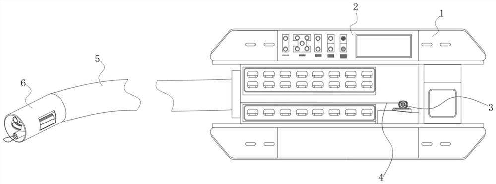
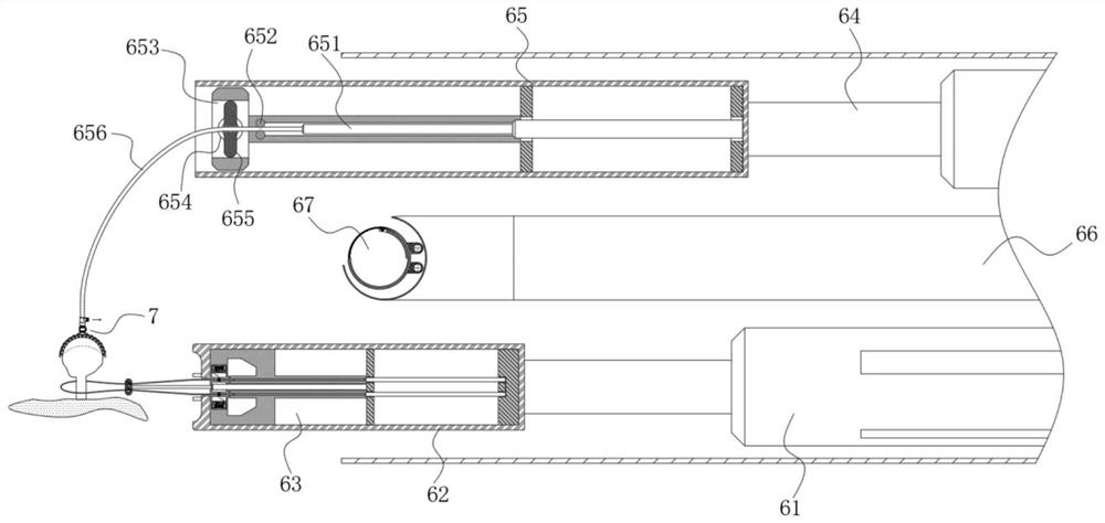
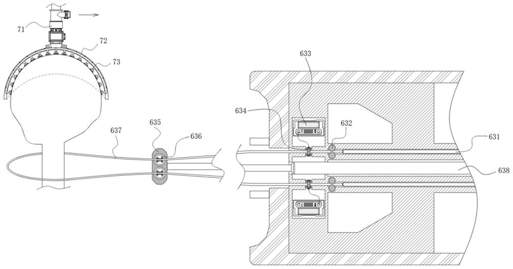
[0036] refer to Figure 1-6, the present invention provides a technical solution: an auxiliary polyp collector for gastrointestinal endoscopy, which includes a storage box 1, a control panel 2, a pull-back winding machine 3, a steel wire rope 4, a gastroscope tube 5, and an extraction device 6. A control panel 2 is installed on the upper end shell of the storage box 1, a polyp storage room is provided in the central shell of the storage box 1, and the gastroscope tube 5 is installed on the left shell wall, and the left end tube of the gastroscope tube 5 is The shell is provided with an extraction device 6, which is provided with an extraction assembly 63, a positioning assembly 65, and a U-shaped delivery tube 66, and the right side corresponding to the two ports on the right side of the U-shaped delivery tube 66 is equipped with Pull back the winding machine 3, and the inside of the U-shaped delivery pipe 66 is equipped with a double-layer guide rod 671 that is set up and dow...


PUM


Abstract

The invention discloses a gastrointestinal endoscope polyp removal auxiliary collector and method. The auxiliary collector comprises a storage box, a control screen, pull-back winding machines, steel wire ropes, a gastroscope tube and a removal device, the control screen is installed on a shell at the upper end of the storage box, a polyp storage chamber is arranged in a central shell of the storage box, the gastroscope tube is installed on the shell wall of the left side of the storage box, a removing device is arranged in a tube shell at the left end of the gastroscope tube, a removing assembly, a positioning assembly and a U-shaped conveying tube are arranged in the removing device, pull-back winding machines are installed on the right sides right corresponding to two ports of the right side of the U-shaped conveying tube,double-layer guide rods which are horizontally arranged up and down are installed in the U-shaped conveying tube, the double-layer guide rods are sleeved with a plurality of sets of collecting boxes arranged at equal intervals, the collecting boxes are tightly connected through the steel wire ropes, the two ends of the collecting boxes are connected with reels in the corresponding pull-back winding machines respectively, and a feeding port is formed in the arc-shaped pipe face of the left side of the U-shaped conveying tube.

Description

technical field [0001] The invention relates to the technical field of medical appliance products, in particular to an auxiliary polyp collector and method for gastrointestinal endoscope extirpation. Background technique [0002] Polyps refer to abnormally growing tissues that protrude from the mucosal surface. They are commonly called polyps before the pathological nature is determined. Generally speaking, polyps are formed due to the growth and aggregation of cells originating from the mucosa. The polyps that grow in the stomach are called gastric polyps, which are common clinical diseases. In clinical practice, most gastric polyps are found incidentally during upper gastrointestinal endoscopy. The vast majority of polyps are benign lesions and are not life-threatening, and only some polyps have a tendency to become cancerous and require active treatment. Moreover, samples of excised polyps need to be preserved for testing to determine the pathological nature of the polyps...

Claims


Application Information

Patent Timeline
IPC IPC(8): A61B18/12A61B18/14A61B90/17
CPCA61B18/12A61B18/14A61B90/17A61B2018/00494A61B2018/00601A61B2018/00982Y02A40/81
Inventor 梅丽娜
Owner HUZHOU MATERNITY & CHILD CARE HOSPITAL
Learn More