Colorectal polyp image recognition method and device and storage medium

A method of identifying, a technique for rectal polyps, used in the medical field

Active Publication Date: 2021-08-20
TIANJIN YUJIN ARTIFICIAL INTELLIGENCE MEDICAL TECH CO LTD
View PDF14 Cites 1 Cited by
  • Summary
  • Abstract
  • Description
  • Claims
  • Application Information

AI Technical Summary

Problems solved by technology

However, at present, it still relies on doctors to judge the results of typing based on experience and visual observation, and there are subjective differences.

Method used


Image

  • Colorectal polyp image recognition method and device and storage medium
  • Colorectal polyp image recognition method and device and storage medium
  • Colorectal polyp image recognition method and device and storage medium

Examples


Embodiment 1

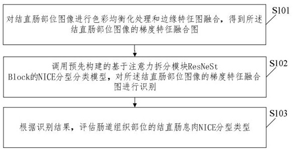
[0050] The embodiment of the present invention provides a method for predicting the result of NICE typing of colorectal polyps, such as figure 1 As shown, the method for predicting the result of NICE typing of colorectal polyps includes:

[0051] S101. Perform color equalization processing and edge feature map fusion on the colorectal part image to obtain a gradient feature fusion map of the colorectal part image;

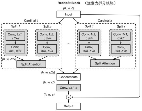
[0052]S102, calling the pre-built NICE classification model based on the attention splitting module ResNeSt Block to identify the gradient feature fusion map of the colorectal part image;

[0053] S103. Evaluate the NICE type of colorectal polyps in the colorectum according to the identification result.

[0054] In the embodiment of the present invention, the color equalization processing and edge feature map fusion processing are performed on the colorectal part image to construct the recognition sample, which can effectively unify and strengthen the image featu...

Embodiment 2

[0110] An embodiment of the present invention provides a colorectal polyp NICE typing result prediction device, the colorectal polyp NICE typing result prediction device includes: a memory, a processor, and a device stored in the memory and operable on the processor computer program;

[0111] When the computer program is executed by the processor, the steps of the method for predicting the result of NICE typing of colorectal polyps as described in any one of the first embodiment are realized.

[0112] Wherein, the device for predicting the result of colorectal polyp NICE typing may be an endoscope detection device, and the storage may be a cloud storage.

Embodiment 3

[0114] An embodiment of the present invention provides a computer-readable storage medium. The computer-readable storage medium stores a colorectal polyp NICE typing result prediction program. When the colorectal polyp NICE typing result prediction program is executed by a processor, The steps of the method for predicting the result of NICE typing of colorectal polyps as described in any one of the first embodiment are realized.


PUM


Abstract

The invention relates to a colorectal polyp image recognition method and device and a storage medium. The colorectal polyp image identification method comprises the steps of performing color equalization processing and edge feature image fusion on a colorectal part image to obtain a gradient feature fusion image of the colorectal part image; calling a pre-constructed classification model of colorectal polyp typing based on an attention splitting module ResNeSt Block, and identifying the gradient feature fusion image of the colorectal part image. According to the method, the recognition sample is constructed by performing color equalization processing and edge feature map fusion processing on the colorectal part image, the image features can be effectively unified and enhanced, feature extraction is performed through the feature extraction network constructed based on ResNeSt Block, the colorectal polyp typing result can be predicted more accurately, and a doctor is assisted in inferring the pathological properties of colorectal polyps.

Description

technical field [0001] The present application relates to the field of medical technology, and in particular to a colorectal polyp image recognition method, device and storage medium. Background technique [0002] Colorectal polyps refer to protruding lesions protruding from the intestinal lumen, and are one of the common intestinal diseases, including adenomatous polyps and non-adenomatous polyps (hamartomatous polyps, metaplastic polyps and inflammatory polyps) , of which adenomatous polyps are precancerous lesions. The morbidity and mortality of colorectal cancer in China are increasing year by year. Most patients are in the advanced stage when they are discovered, and the 5-year survival rate is less than 50%. Colonoscopy is the most intuitive and effective way to find lesions, which can improve the detection of early intestinal cancer and reduce mortality. Electronic chromoendoscopy is an important progress in the field of digestive endoscopy in recent years, such as ...

Claims


Application Information

Patent Timeline
Patent Type & Authority Applications(China)
IPC IPC(8): G06T7/00G06K9/46G06K9/62G06N3/04
CPCG06T7/0012G06T2207/10068G06T2207/30032G06V10/44G06N3/045G06F18/253G06F18/214
Inventor 李佳昕王玉峰
Owner TIANJIN YUJIN ARTIFICIAL INTELLIGENCE MEDICAL TECH CO LTD
Why Eureka
Try Eureka