Cancer auxiliary analysis system and device based on he-stained pathological images
An auxiliary analysis and pathological image technology, applied in the field of medical imaging, can solve the problem of inability to accurately segment the cytoplasm, and achieve the effect of effective network model parameters, ensuring segmentation accuracy, and good segmentation accuracy and accuracy.
- Summary
- Abstract
- Description
- Claims
- Application Information
AI Technical Summary
Problems solved by technology
Method used
Image

Examples
specific Embodiment approach 1
[0049]The existing cell segmentation methods cannot accurately segment the cytoplasm. On the one hand, the existing segmentation network model itself cannot accurately segment the cytoplasm, that is, for complex and difficult-to-divide images, the segmentation result of the network model itself is not enough. There is a problem of inaccuracy; on the other hand, due to the current HE staining, the layers are not clear enough, the distinction between the nucleus and the cytoplasm is not obvious, the layers of the cytoplasm and the extracellular space are less clear, and the distinction is even less obvious, which further reduces the staining image of the neural network. Handling accuracy.
[0050] This embodiment is a cancer auxiliary analysis system based on HE-stained pathological images, including:
[0051] The dyeing section image acquisition module is used to obtain the HE-stained dyed section image, and segment the image into image blocks;
[0052] The cell nucleus segmen...
specific Embodiment approach 2
[0089] This embodiment is a cancer auxiliary analysis system based on HE-stained pathological images, further comprising:
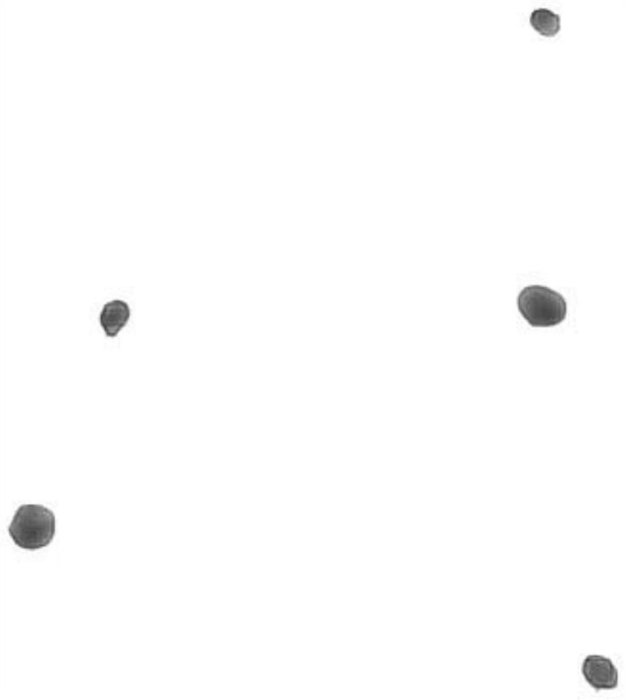
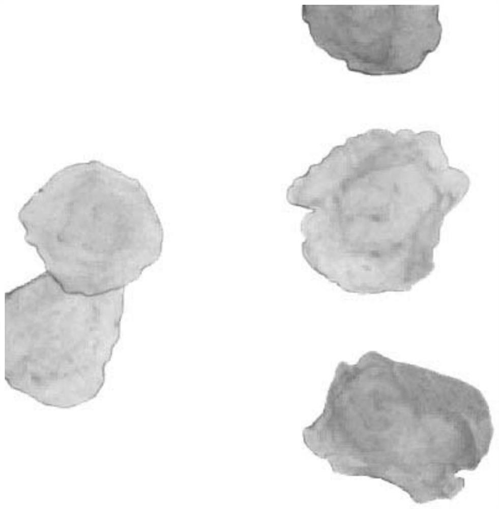
[0090] The overall cell unit determination module, for the image block corresponding to the stained slice image, maps the segmentation result of the cytoplasmic segmentation module to the corresponding image block, and maps the segmentation result of the nucleus segmentation module to the same image block, and finally forms the stained slice. A segmented image of an image, such as Figure 4 shown.
[0091] In fact, the results of the cytoplasmic segmentation module are mapped to the corresponding image blocks, while the results of the nucleus segmentation module are mapped to the same image block. Sometimes, multiple nuclei are mapped in the same cytoplasmic divided area. As long as the situation occurs when cells accumulate or become cancerous, such effects will not affect the auxiliary analysis of cancer. You can add reference factors for the arrangeme...
specific Embodiment approach 3
[0092] This embodiment is a cancer auxiliary analysis system based on HE-stained pathological images, further comprising:
[0093] The cancer auxiliary analysis module is used to identify and classify cancerous cells based on the results of the cell whole unit determination module using the expert database. The identification and classification process is carried out by means of an expert database, which stores the judgment rules of cancerous cells, and the judgment rules of cancerous cells are the morphological characteristics of cancerous cells determined by experts based on the big data of pathological images, such as the arrangement of nuclei or cells The state (whether disordered, agglomerated into pieces, etc.), the size of the nucleus (the size of each nucleus, and whether multiple nuclei are different in size, etc.), the shape of the nucleus, etc., this embodiment is characterized in that it can also include cytoplasm related morphological features, such as nucleocytop...
PUM
 Abstract
Description
Claims
Application Information
 - R&D
- Intellectual Property
- Life Sciences
- Materials
- Tech Scout
- Unparalleled Data Quality
- Higher Quality Content
- 60% Fewer Hallucinations
Browse by: Latest US Patents, China's latest patents, Technical Efficacy Thesaurus, Application Domain, Technology Topic, Popular Technical Reports.
© 2025 PatSnap. All rights reserved.Legal|Privacy policy|Modern Slavery Act Transparency Statement|Sitemap|About US| Contact US: help@patsnap.com