Elastic imaging method and device, electronic equipment and storage medium

An elastic imaging and elastic detection technology, applied in medical science, diagnosis, application, etc., can solve problems such as poor quality of ultrasonic imaging and inaccurate detection results, so as to improve the accuracy, improve the quality of ultrasonic imaging, and eliminate poor test positions Effect

Pending Publication Date: 2021-07-09
WUXI HISKY MEDICAL TECH +1
View PDF3 Cites 2 Cited by
  • Summary
  • Abstract
  • Description
  • Claims
  • Application Information

AI Technical Summary

Problems solved by technology

[0006] The present application provides an elastic imaging method, device, electronic equipment and storage medium to solve the problem of inaccurate detection results due to the poor quality of ultrasonic imaging in the prior art

Method used


Image

  • Elastic imaging method and device, electronic equipment and storage medium
  • Elastic imaging method and device, electronic equipment and storage medium
  • Elastic imaging method and device, electronic equipment and storage medium

Examples


Embodiment Construction

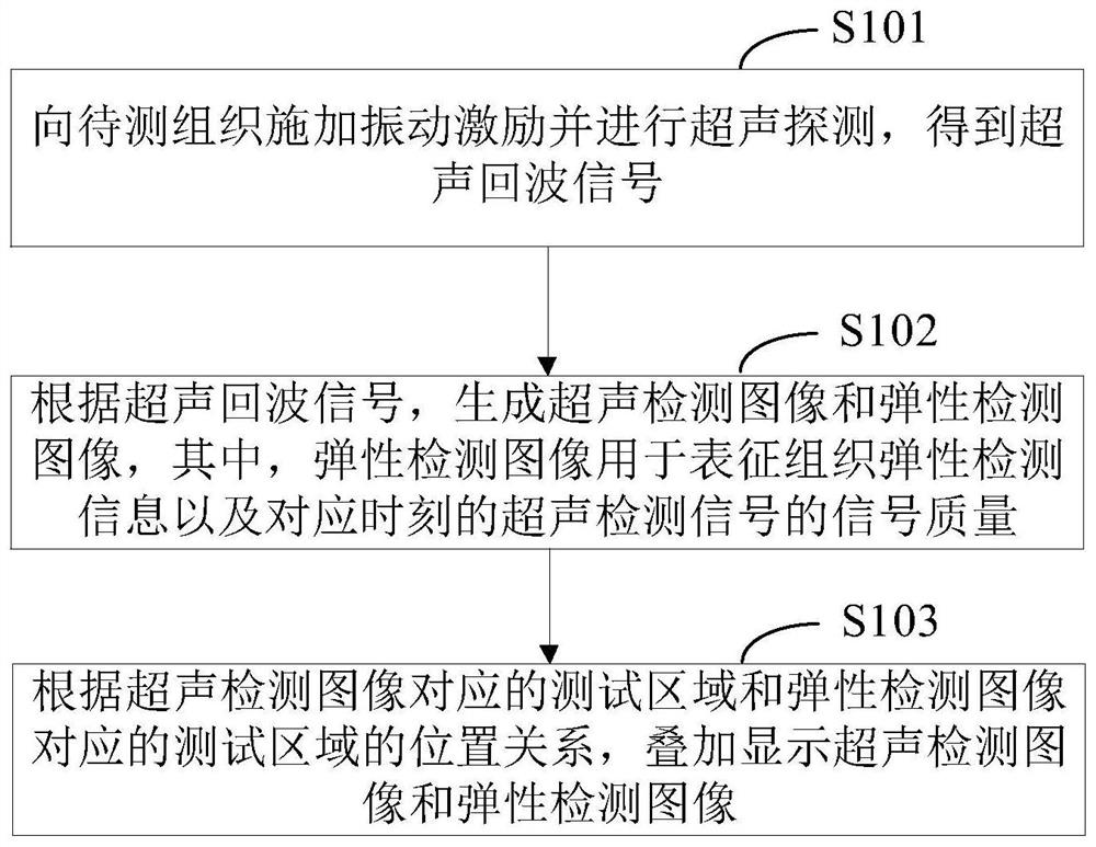
[0033] Reference will now be made in detail to the exemplary embodiments, examples of which are illustrated in the accompanying drawings. When the following description refers to the accompanying drawings, the same numerals in different drawings refer to the same or similar elements unless otherwise indicated. The implementations described in the following exemplary embodiments do not represent all implementations consistent with this application. Rather, they are merely examples of apparatuses and methods consistent with aspects of the present application as recited in the appended claims.

[0034] The application scenarios of the embodiments of the present application are explained below:

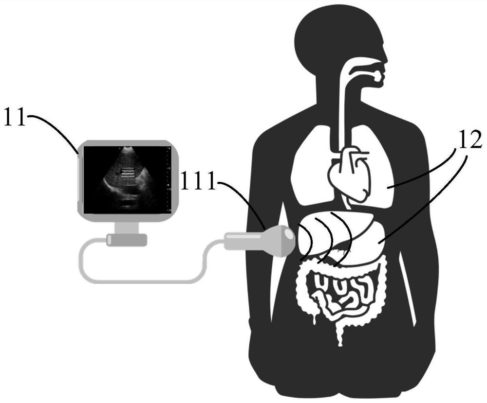
[0035] figure 1 An application scene diagram of the elastography method provided by the embodiment of this application, such as figure 1 As shown, the elastography method provided by the embodiment of the present application is applied to an electronic device, specifically, for example...


PUM


Abstract

The embodiment of the invention provides an elastic imaging method and device, electronic equipment and a storage medium. The method comprises the steps: applying vibration excitation to a to-be-detected tissue, and carrying out the ultrasonic detection, thereby obtaining an ultrasonic echo signal; according to the ultrasonic echo signal, an ultrasonic detection image and an elasticity detection image are generated, and the elasticity detection image is used for representing tissue elasticity detection information and the signal quality of the ultrasonic detection signal at the corresponding moment; according to the position relation between the test region corresponding to the ultrasonic detection image and the test region corresponding to the elasticity detection image, the ultrasonic detection image and the elasticity detection image are displayed in an overlapped mode, and after the ultrasonic detection image is generated, the elasticity detection image is displayed on the ultrasonic detection image in an overlapped mode. The imaging quality of the ultrasonic detection image is represented by using the elastic detection image, so that an operator can quickly control the quality of the detection result, the influence caused by the reasons such as poor test position and breath of the detected human body is eliminated, the ultrasonic imaging quality is improved, and the accuracy of the detection result is improved.

Description

technical field [0001] The present application relates to the technical field of medical equipment, in particular to an elastic imaging method, device, electronic equipment and storage medium. Background technique [0002] This section is intended to provide a background or context to the implementations of the application that are recited in the claims. The descriptions herein are not admitted to be prior art by inclusion in this section. [0003] Ultrasound imaging technology is widely used in clinical detection because of its advantages of real-time, cheap, non-invasive and non-ionizing radiation. By obtaining the physiological structure information of biological tissue, it can realize the detection of biological tissue lesions. However, in the early lesions of biological tissues, the traditional B-ultrasound and other ultrasonic imaging methods are not sensitive to the early lesions of biological tissues because their structure does not change significantly. Elastic im...

Claims


Application Information

Patent Timeline
IPC IPC(8): A61B8/00
CPCA61B8/485A61B8/5215A61B8/5261
Inventor 何琼邵金华孙锦贾继东尤红欧晓娟
Owner WUXI HISKY MEDICAL TECH
Why Eureka
Try Eureka