
Liver tumor image sample augmentation method based on generative adversarial network

A liver tumor and image sample technology, applied in the field of liver tumor image sample augmentation, to achieve the effect of enriching reality and increasing variability

Pending Publication Date: 2021-06-15
北京精诊医疗科技有限公司
View PDF3 Cites 2 Cited by
  • Summary
  • Abstract
  • Description
  • Claims
  • Application Information

AI Technical Summary

Problems solved by technology

However, there is a large error between the sample image obtained by performing a simple transformation operation on the image and the actual image.

Method used


Image

  • Liver tumor image sample augmentation method based on generative adversarial network
  • Liver tumor image sample augmentation method based on generative adversarial network

Examples


Embodiment Construction

[0020] Below in conjunction with accompanying drawing and embodiment, technical solution of the present invention is described further:

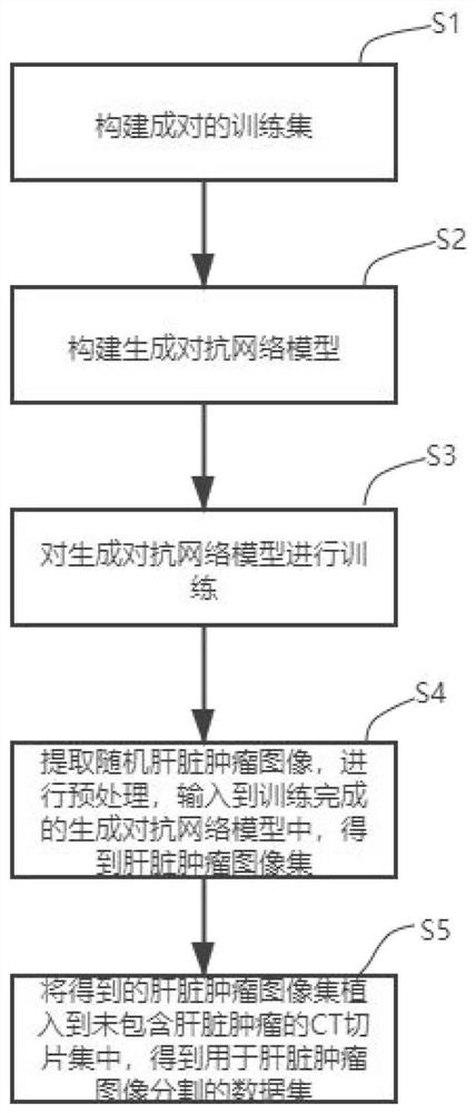
[0021] This embodiment provides a method for augmenting liver tumor image samples based on generative adversarial networks, such as figure 1 shown, including:

[0022] S1, construct a paired training set;

[0023] The steps include acquiring a CT slice containing a liver tumor, extracting a tumor image in the CT slice containing a liver tumor, obtaining an original tumor image, and performing preprocessing on the original tumor image to generate a tumor image;

[0024] In the embodiment of this application, CT slices with liver tumors are obtained from the Liver Tumor Segmentation Challenge Dataset (LiTS) as the original tumor images, and the corresponding masks are set according to the tumor size in the CT slices to ensure that the set mask The phantom can cover the tumor in the CT slice. And the tumor mask image, that is, the original t...


PUM


Abstract

The invention discloses a liver tumor image sample augmentation method based on a generative adversarial network, and the method comprises the following steps: firstly constructing a paired training set, then constructing a generative adversarial network model which comprises a generator and a discriminator, and training the generative adversarial network model; extracting a random liver tumor image from the CT slice data set of the liver tumor, preprocessing the random liver tumor image, inputting the random liver tumor image into the trained generative adversarial network model to obtain a liver tumor image set, finally implanting the obtained liver tumor image set into the CT slice set not containing the liver tumor, and obtaining a data set for liver tumor image segmentation. According to the invention, the random liver tumor image can be generated through the generative adversarial network model, so that the augmentation of the liver tumor data set is realized, the variability of the liver tumor is improved, and a rich and real liver tumor slice data set is created for the liver slice.

Description

technical field [0001] The invention belongs to the field of medical image processing, and in particular relates to a liver tumor image sample augmentation method based on a generative confrontation network. Background technique [0002] With the advancement of science and technology, medical imaging technology has made great progress. Image segmentation is an indispensable means of extracting quantitative information of special tissues in medical images. In order to accurately distinguish normal tissue structures and abnormal lesions in medical images, it is necessary to Segmentation of medical images is a key step in medical image processing. [0003] Since a large number of parameters need to be tuned in image segmentation model training, there is a high demand for the number of training samples. Therefore, data augmentation of training data has become one of the main means of applying deep learning technology. This method mainly performs transformation operations on ima...

Claims


Application Information

Patent Timeline
IPC IPC(8): G06T7/00G06T7/11G06K9/62
CPCG06T7/0012G06T7/11G06T2207/10081G06T2207/20081G06T2207/30056G06T2207/30096G06F18/214
Inventor 王博赵威申建虎张伟徐正清
Owner 北京精诊医疗科技有限公司
Learn More