
Detection probe for SNP site detection, detection method and application of detection probe

A technology for detecting probes and sites, applied in biochemical equipment and methods, recombinant DNA technology, microbial measurement/inspection, etc., to achieve the effects of fast hole passing, improved detection resolution, and reduced translocation speed

Pending Publication Date: 2021-06-11
NANJING UNIV OF POSTS & TELECOMM
View PDF4 Cites 0 Cited by
  • Summary
  • Abstract
  • Description
  • Claims
  • Application Information

AI Technical Summary

Problems solved by technology

However, the high sensitivity of nanopore signal detection requires that the size of the nanopore is similar to that of the detection molecule. In order to realize the recognition of a single base, an ultra-thin solid nanopore of sub-2nm is also required. Nanopore signal acquisition and amplification devices have certain challenges

Method used


Image

  • Detection probe for SNP site detection, detection method and application of detection probe
  • Detection probe for SNP site detection, detection method and application of detection probe
  • Detection probe for SNP site detection, detection method and application of detection probe

Examples


Embodiment 1

[0061] Embodiment 1: the preparation of detection probe

[0062] Step 1: Take an appropriate amount of the first nucleic acid probe (SEQ ID NO.1) and gold nanospheres with a particle size of about 5nm, mix the DNA solution and the gold nanospheres according to the ratio range (molar ratio) of 1:10, shake well, Cultivate overnight in an incubator at 25°C and 300 rpm.

[0063] Step 2: Take the PBS buffer solution with a volume of about 10% of the mixed solution in step (1), so that the PBS concentration in the final solution is about 0.1mol / L. The PBS buffer solution is divided into 5 times with an interval of 20 minutes each time Add, fully shake and shake well after each addition, after all the addition, incubate overnight in an incubator at 25°C and 300rpm:

[0064] Step 3: Place the sample in a high-speed temperature-controlled centrifuge, conditions: 15,000rpm, 25°C, after high-speed centrifugation, a precipitate appears at the bottom, that is, gold nanospheres modified wi...

Embodiment 2

[0066] Embodiment 2: nanopore sensor detection

[0067] In this embodiment, the present invention selects the wild-type and mutant DNA fragments in the p53 gene as detection objects.

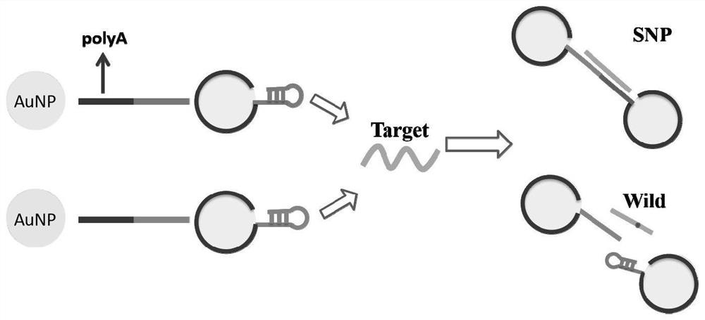
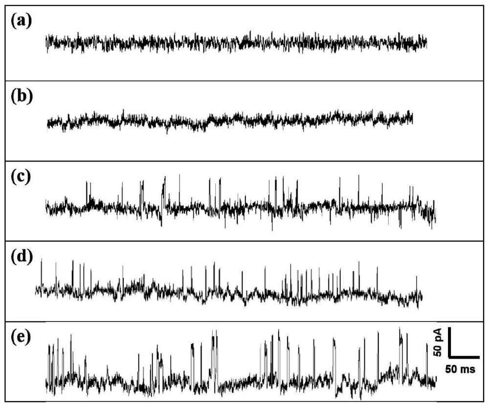
[0068] Step 1: Take the two probe samples prepared in Example 1, mix the two probes in a ratio of 1:1, and shake well. Then react with wild-type (SEQ ID NO.3) and SNP site mutant (SEQ ID NO.4) target molecules respectively according to the molar ratio of 1:100, shake well, and incubate in an incubator at 25°C and 300rpm overnight.

[0069] Step 2: Place the mixture of the probe sample prepared in step 1, wild type (SEQ ID NO.3) and SNP site mutant (SEQ ID NO.4) in a temperature-controlled centrifuge (25°C, 12000r .p.m), remove the supernatant, precipitate out, redissolve with PBS buffer solution with a concentration of 0.1mol / L, and store in a refrigerator at 4°C for nanopore detection.

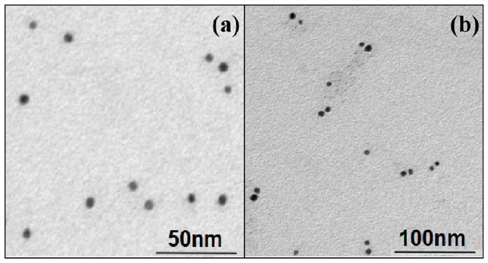
[0070] Step 3: Observing the complex of the prepared detection probe and the target molecule with a 40nm ...


PUM

PropertyMeasurementUnit
diameteraaaaaaaaaa
diameteraaaaaaaaaa
particle diameteraaaaaaaaaa

Abstract

The invention discloses a detection probe for SNP (Single Nucleotide Polymorphism) site detection, a detection method and application of the detection probe. The detection probe comprises a first detection probe and a second detection probe; the first detection probe comprises a gold nanosphere and a first nucleic acid probe modifying the surface of the gold nanosphere, the first nucleic acid probe comprises an anchoring chain connected with the gold nanosphere and a first recognition chain used for capturing SNP target molecules, and the first recognition chain is of a hairpin structure; and the second detection probe comprises a gold nanosphere and a second nucleic acid probe modifying the surface of the gold nanosphere, the second nucleic acid probe comprises an anchoring chain connected with the gold nanosphere and a second recognition chain used for capturing SNP target molecules, and the second recognition chain is of a hairpin structure. The detection method provided by the invention overcomes the defect of insufficient resolution of solid nanopores in DNA single base mutation detection, realizes high-sensitivity detection of the solid nanopore on the SNP site, and expands the application of the solid nanopore in the field of single molecule detection.

Description

technical field [0001] The invention relates to the field of nanopore sensor detection, in particular to a detection probe for SNP site detection, a detection method and an application thereof. Background technique [0002] Single nucleotide polymorphisms (Single nucleotide polymorphisms, SNP) refers to the polymorphism of nucleic acid sequence caused by the change of a single nucleotide base in the gene sequence, including single base conversion, insertion and deletion. SNPs are widely distributed in the genome, and studies have shown that they appear once every 300 base pairs in the human genome. The large number of SNP loci gives people the opportunity to discover genomic mutations associated with various diseases, including tumors. It is easier to discover disease-associated gene mutations through SNPs than through family genetic maps; some SNPs do not directly lead to the expression of disease genes, but because they are adjacent to certain disease genes, they become i...

Claims


Application Information

Patent Timeline
Patent Type & Authority Applications(China)
IPC IPC(8): C12Q1/6886C12Q1/6825C12Q1/6827C12N15/11
CPCC12Q1/6886C12Q1/6825C12Q1/6827C12Q2600/156C12Q2525/173C12Q2525/301C12Q2563/137C12Q2563/155C12Q2565/519C12Q2565/631C12Q2565/607Y02A50/30
Inventor 翁丽星武灵芝叶媛席国豪严馨曾祥杰
Owner NANJING UNIV OF POSTS & TELECOMM
Learn More