
Magnetic anchoring pulmonary nodule positioning device for thoracoscopic surgery

A positioning device and thoracoscopic technology, applied in the field of medical equipment, can solve problems such as displacement or falling off, poor positioning effect, and unfavorable recovery of patients

Pending Publication Date: 2021-05-11
NANTONG TUMOR HOSPITAL
View PDF0 Cites 0 Cited by
  • Summary
  • Abstract
  • Description
  • Claims
  • Application Information

AI Technical Summary

Problems solved by technology

However, for pulmonary nodules with a subpleural depth of more than 5 mm and a diameter of less than 10 mm, conventional methods such as vision, finger palpation, or surgical instrument sliding cannot be used to accurately locate pulmonary nodules during thoracoscopic surgery, which may lead to prolonged operation time and excessive resection of normal lung tissue etc., which in turn affects the curative effect of surgery, increases postoperative complications, is not conducive to the recovery of patients, and increases the possibility of conversion to thoracotomy
[0003] In order to optimize the application of thoracoscopy in pulmonary nodule resection, a series of pulmonary nodule localization techniques have been explored clinically in recent years, such as puncture localization technology, image-mediated localization technology, electromagnetic navigation bronchoscopy localization, etc. The clamping position is displaced or falls off, and the positioning effect is not good, and some need a pair of positioning structures for positioning, so two puncture processes are required, which is not conducive to postoperative recovery for patients

Method used


Image

  • Magnetic anchoring pulmonary nodule positioning device for thoracoscopic surgery
  • Magnetic anchoring pulmonary nodule positioning device for thoracoscopic surgery
  • Magnetic anchoring pulmonary nodule positioning device for thoracoscopic surgery

Examples


Embodiment 1

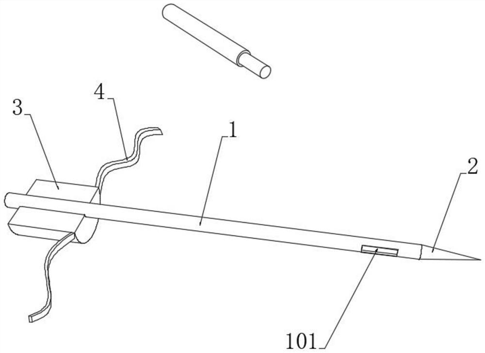
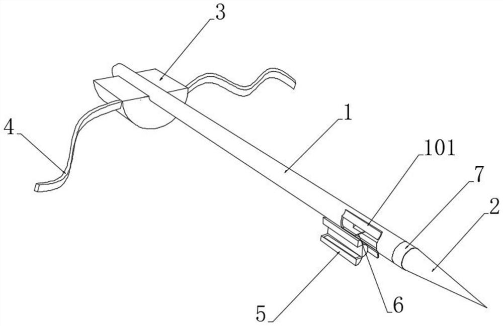
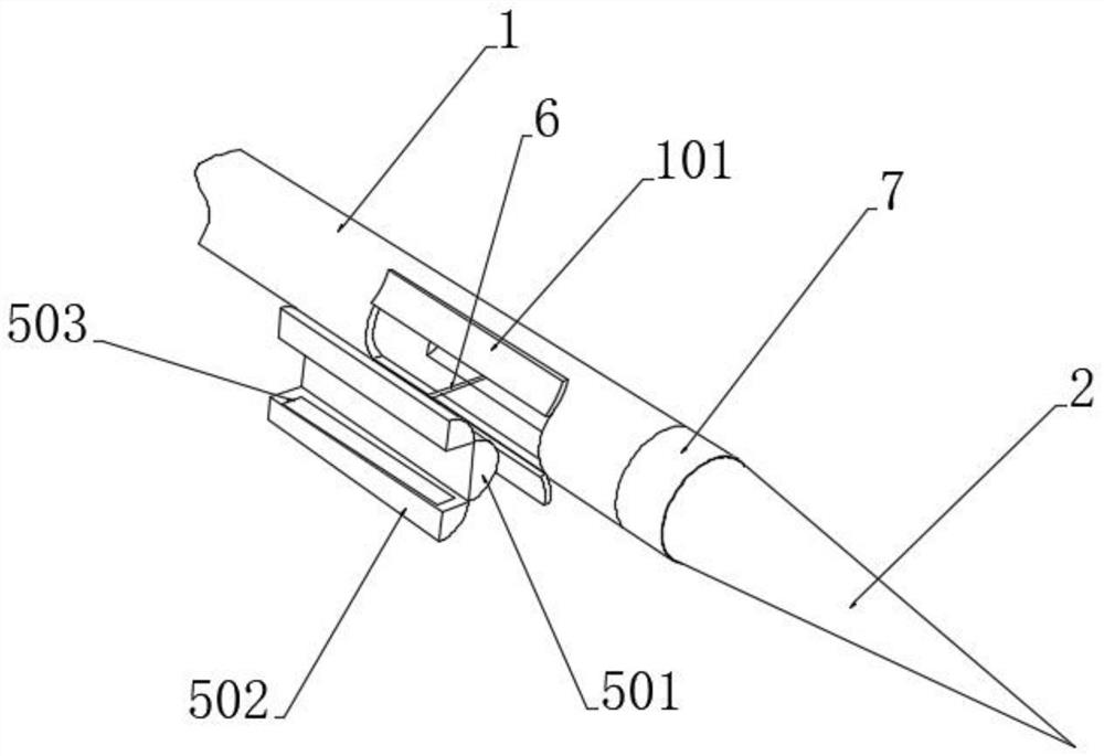
[0039] see figure 1 , a magnetically anchored pulmonary nodule positioning device for thoracoscopic surgery, comprising a puncture needle tube 1, a positioning pad 3 and a magnetic suction rod, the front end of the puncture needle tube 1 is provided with a puncture needle 2, and the upper end of the positioning pad 3 is provided with a The sliding cavity matched with the puncture needle tube 1, the inner end surface of the positioning pad 3 is covered with a flexible layer, and both sides of the inner end of the positioning pad 3 are provided with attachment straps 4, and an adhesive layer is provided on the attachment strap 4 , during medical use, the positioning pad 3 is attached to the outer surface of the body of the patient's lung tissue to be observed, and a pair of attachment straps 4 are used to position the positioning pad 3, and the puncture needle tube 1 is slidably connected to the positioning pad 3 , it is easy for medical staff to grasp the puncture angle more ac...


PUM


Abstract

The invention discloses a magnetic anchoring pulmonary nodule positioning device for a thoracoscopic surgery, and belongs to the technical field of medical instruments. A targeted positioning body is placed in a puncture needle tube, a micro-driving body and a pair of arc-shaped magnetic closing layers limit the targeted positioning body, when the puncture needle tube penetrates into lung tissue, the targeted positioning body is pushed outwards to be placed at the target nodule by utilizing the micro-driving body, one pair of positioning flaps rotationally connected to a magnetized elliptic block is overturned outwards under the reset action of torsional elasticity to form a clamping space, and one pair of extending clamping surfaces attract each other to move through magnetic attraction force, so that positioning of the target nodule is completed; two positioning structures do not need to be placed on the target nodule, so that the operation is relatively easy and convenient; and during surgical operation, a magnetic suction rod is clamped through surgical forceps to penetrate into the lung tissue and slightly slides on the surface of the lung tissue, and the surface of the pulmonary membrane is attracted to lift a bulge, namely the position where the target nodule is located, so that the excision operation can be conveniently conducted on the lifted bulge.

Description

technical field [0001] The invention relates to the technical field of medical instruments, in particular to a magnetically anchored pulmonary nodule positioning device for thoracoscopic surgery. Background technique [0002] As the main technical method for the diagnosis and treatment of pulmonary nodules, thoracoscopic technology has the advantages of less trauma, faster recovery, and fewer surgical complications, and is widely used in clinical practice. However, for pulmonary nodules with a subpleural depth of more than 5 mm and a diameter of less than 10 mm, conventional methods such as vision, finger palpation, or surgical instrument sliding cannot be accurately located during thoracoscopic surgery, which may lead to prolonged operation time and excessive resection of normal lung tissue etc., which in turn affects the curative effect of the operation, increases postoperative complications, is not conducive to the recovery of patients, and increases the possibility of co...

Claims


Application Information

Patent Timeline
IPC IPC(8): A61B90/11A61B34/20A61B17/34
CPCA61B17/3403A61B34/20A61B90/11A61B2034/2051
Inventor 樊怿辉陈赛华施民新
Owner NANTONG TUMOR HOSPITAL
Learn More