Pulmonary nodule interactive segmentation method based on medical image

A technology for medical imaging and pulmonary nodules, applied in the field of medical image processing, can solve time-consuming and labor-intensive problems

Pending Publication Date: 2021-03-30
睿佳(武汉)软件科技有限公司
View PDF1 Cites 0 Cited by
  • Summary
  • Abstract
  • Description
  • Claims
  • Application Information

AI Technical Summary

Problems solved by technology

In the process of clinical diagnosis, doctors need to screen nodules one by one in hundreds of CT slices, which not only greatly depends on the doctor's clinical experience, but also is extremely time-consuming and labor-intensive

Method used


Image

  • Pulmonary nodule interactive segmentation method based on medical image
  • Pulmonary nodule interactive segmentation method based on medical image
  • Pulmonary nodule interactive segmentation method based on medical image

Examples


Embodiment

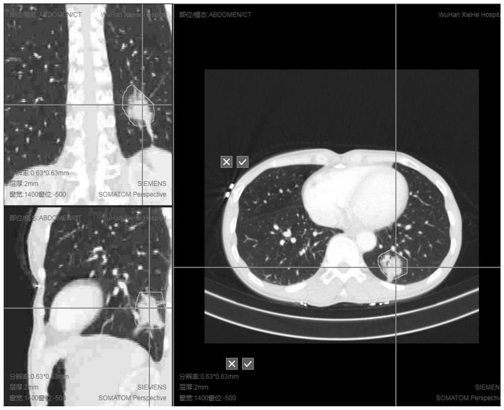
[0162] image 3 ) ROI is manually drawn, and the grid area on the map is the manually drawn area. After completion, click "√";

[0163] Such as image 3 As shown, multi-planar reconstruction was performed on the lung CT, and the lung nodules were roughly outlined on the three axial planes of the transverse plane, coronal plane, and sagittal plane, as shown in the grid area in the figure. Click "√" to confirm after drawing.

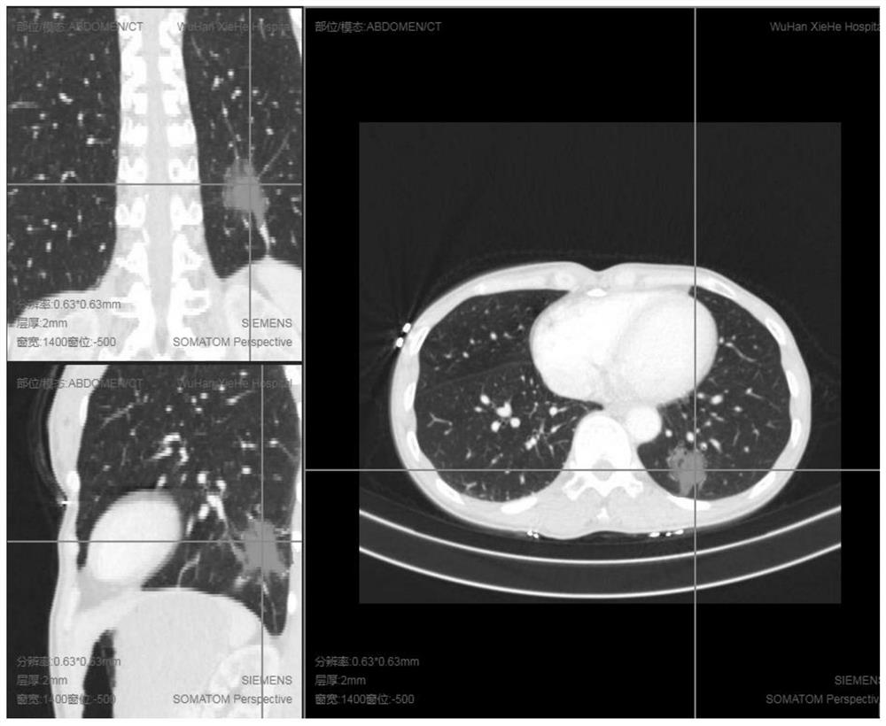
[0164] After the sketch is completed, proceed as figure 2 In the algorithm flow shown, the automatic segmentation result is obtained, that is, the maximum connected region. Automatically segment results such as Figure 4 shown.

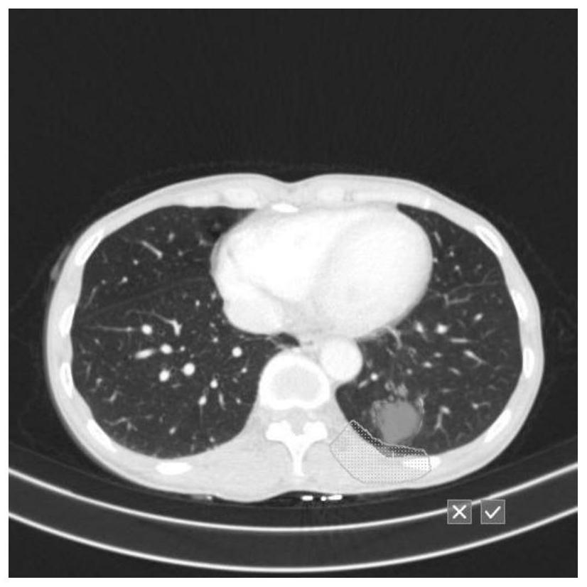
[0165] The results of automatic segmentation can be further adjusted, such as Figure 5 shown. Figure 3-6 In order to manually outline the single-layer subtraction area and its result (the area is the area that needs to be subtracted), Figure 7-10 is the truncated area obtained by intermediate interpolation between the ...


PUM


Abstract

The invention discloses a pulmonary nodule interactive segmentation method based on a medical image. The method comprises the following steps: extracting a focus area according to a real nodule and aground glass nodule, wherein the segmentation algorithm is a semi-automatic segmentation algorithm, the lung real nodule segmentation method needs artificial participation to delineate a nodule region, and the lung frosted glass nodule segmentation method is automatic segmentation; if modification is needed after segmentation is completed, selecting two-dimensional modification or three-dimensional modification according to requirements to obtain an accurate segmentation result. Therefore, the rapid segmentation of pulmonary nodules is realized for further diagnosis and analysis.

Description

technical field [0001] The invention belongs to the technical field of medical image processing, in particular to an interactive segmentation method of pulmonary nodules based on medical images. Background technique [0002] The situation of lung nodule screening for early prevention and control of lung cancer is still grim. There are many people suffering from lung cancer in my country, the incidence rate is high, and the medical expenses are large. According to the order of incidence, lung cancer accounts for 20.3% of all cancers, ranking first in the incidence of malignant tumors. According to the "National Cancer Report 2019" issued by the National Cancer Quality Control Center, the death rate of malignant tumors is as high as 23.91% of all residents' deaths, and the incidence and death of malignant tumors have continued to rise in the past ten years. More than 220 billion. In the process of clinical diagnosis, doctors need to screen nodules one by one in hundreds of ...

Claims


Application Information

Patent Timeline
Patent Type & Authority Applications(China)
IPC IPC(8): G06T7/11G06T7/136G06T7/194G06T5/30G06T7/187
CPCG06T7/11G06T7/136G06T7/194G06T5/30G06T7/187G06T2207/10081G06T2207/20104G06T2207/20132G06T2207/30064
Inventor 袁戎石姝玥艾鸽
Owner 睿佳(武汉)软件科技有限公司
Why Eureka
Try Eureka