
Definition circle HSV color space based medical image segmentation method and cancer cell identification method

A color space, medical image technology, applied in the field of cancer cell recognition and medical image segmentation, can solve the problem of not considering the influence of saturation and illumination value, and achieve the effect of high segmentation accuracy

Active Publication Date: 2015-12-09
BEIJING INSTITUTE OF TECHNOLOGYGY
View PDF4 Cites 22 Cited by
  • Summary
  • Abstract
  • Description
  • Claims
  • Application Information

AI Technical Summary

Problems solved by technology

But often these segmentation methods for HSV space still have certain errors, because this type of method is only for the image segmentation of the "H" channel, and does not consider the saturation and illumination values ​​"S" and "V". channel effect

Method used


Image

  • Definition circle HSV color space based medical image segmentation method and cancer cell identification method
  • Definition circle HSV color space based medical image segmentation method and cancer cell identification method
  • Definition circle HSV color space based medical image segmentation method and cancer cell identification method

Examples


Embodiment Construction

[0031] The present invention will be described in detail below in conjunction with the accompanying drawings and specific embodiments.

[0032] The present invention proposes a brand-new segmentation method based on real colors, which uses a defined circle to analyze in the HSV three-channel space and maps back to the RGB three-channel space to perform segmentation for a specific color. This method meets the high-precision segmentation and analysis requirements of medical images, and has good adaptability to the layering and gradient of real colors.

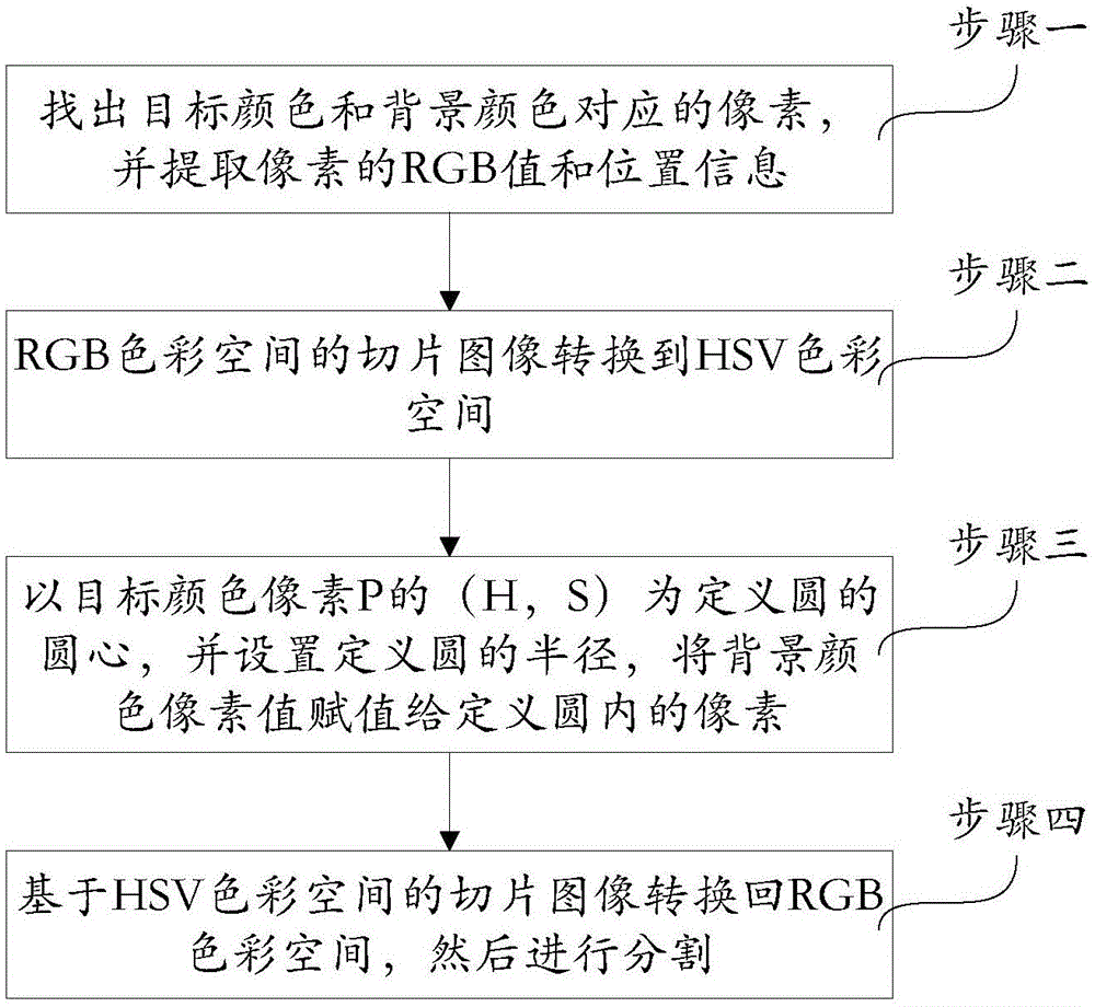
[0033] Such as figure 1 Shown, a kind of medical image segmentation method based on definition circle HSV color space of the present invention, the specific process of this method is:

[0034]Step 1. Find a pixel P corresponding to the target color in the slice image in the RGB color space, and extract the RGB value and position information of the pixel P; find a pixel Q corresponding to the background color in the slice image i...


PUM


Abstract

The invention provides a definition circle HSV color space based medical image segmentation method and a cancer cell identification method. The specific process comprises: step 1, finding out RGB values and position information of a slice image target color pixel P and a background color pixel Q in an RGB color space; step 2, converting an RGB color space based slice image to the HSV color space to obtain an HSV color space based image; step 3, according to position information of the stored pixel P, taking (H,S) corresponding to the pixel P as circle center coordinates of a definition circle, and setting a radius of the definition circle; according to the position information of the pixel Q, extracting H, S and V values corresponding to the pixel Q, assigning the values to all pixels in the definition circle, and removing a target color; and step 4. converting the HSV color space based slice image subjected to removal of the target color back to the RGB color space, and then segmenting the slice image subjected to removal of the target color. By utilizing the definition circle HSV color space based medical image segmentation method and the cancer cell identification method, an extremely accurate segmentation result can be obtained.

Description

technical field [0001] The invention belongs to the technical field of image segmentation, and in particular relates to a medical image segmentation method and a cancer cell identification method based on a defined circular HSV (hue, saturation, illumination) color space. Background technique [0002] Image segmentation is a very widely used technology in the field of image processing, such as video tracking, remote sensing image analysis, medical image analysis, computer vision and other fields will involve image segmentation issues. At present, there are many relatively mature methods in the field of image segmentation. Generally speaking, there are three main entry points for image segmentation: first, the segmentation method based on the gray level of the image target; and second, the segmentation method based on the topology of the image target; The last is the segmentation method based on the image target color. [0003] The classic algorithm based on the target gray ...

Claims


Application Information

Patent Timeline
Patent Type & Authority Applications(China)
IPC IPC(8): G06T7/00G06T7/40
CPCG06T2207/20152G06T2207/30024
Inventor 陈禾庄胤马龙毕福昆陈亮龙腾
Owner BEIJING INSTITUTE OF TECHNOLOGYGY
Learn More