Method for establishing simulated three-dimensional intracranial vascular stenosis analysis model

A technology for intracranial blood vessels and three-dimensional models, applied in the field of image processing, can solve problems such as location and analysis of unfavorable intracranial blood vessel lesion areas, and achieve the effects of facilitating the location and display of stenotic lesion areas, facilitating intuitive observation, and improving registration accuracy.

Inactive Publication Date: 2021-03-16
XIAN CREATION KEJI CO LTD
View PDF0 Cites 2 Cited by
  • Summary
  • Abstract
  • Description
  • Claims
  • Application Information

AI Technical Summary

Problems solved by technology

[0005] However, from the above magnetic resonance images, it is impossible to obtain intuitive and fast analysis data on the degree of intracranial vascular stenosis, which is not conducive to the clinical positioning and analysis of intracranial vascular lesion regions.

Method used


Image

  • Method for establishing simulated three-dimensional intracranial vascular stenosis analysis model
  • Method for establishing simulated three-dimensional intracranial vascular stenosis analysis model
  • Method for establishing simulated three-dimensional intracranial vascular stenosis analysis model

Examples


Embodiment approach

[0153] An optional implementation of this step includes:

[0154] Perform gray-scale linear transformation on each registered bright blood image to obtain K contrast-enhanced bright blood images;

[0155] In the embodiment of the present invention, according to the characteristic that the blood in the bright blood image shows high signal, while the surrounding brain tissue shows low signal, grayscale linear transformation is performed on the bright blood image after registration, and the gray scale range of the image is adjusted to achieve the purpose of improving image contrast. .

[0156] For example, the grayscale linear transformation and parameter settings used in a bright blood image after registration are as follows: Figure 12 as shown, Figure 12 It is a schematic diagram of grayscale linear transformation and parameter setting provided by the embodiment of the present invention. use Figure 12 The gray scale linear transformation shown can extend the smaller rang...


PUM


Abstract

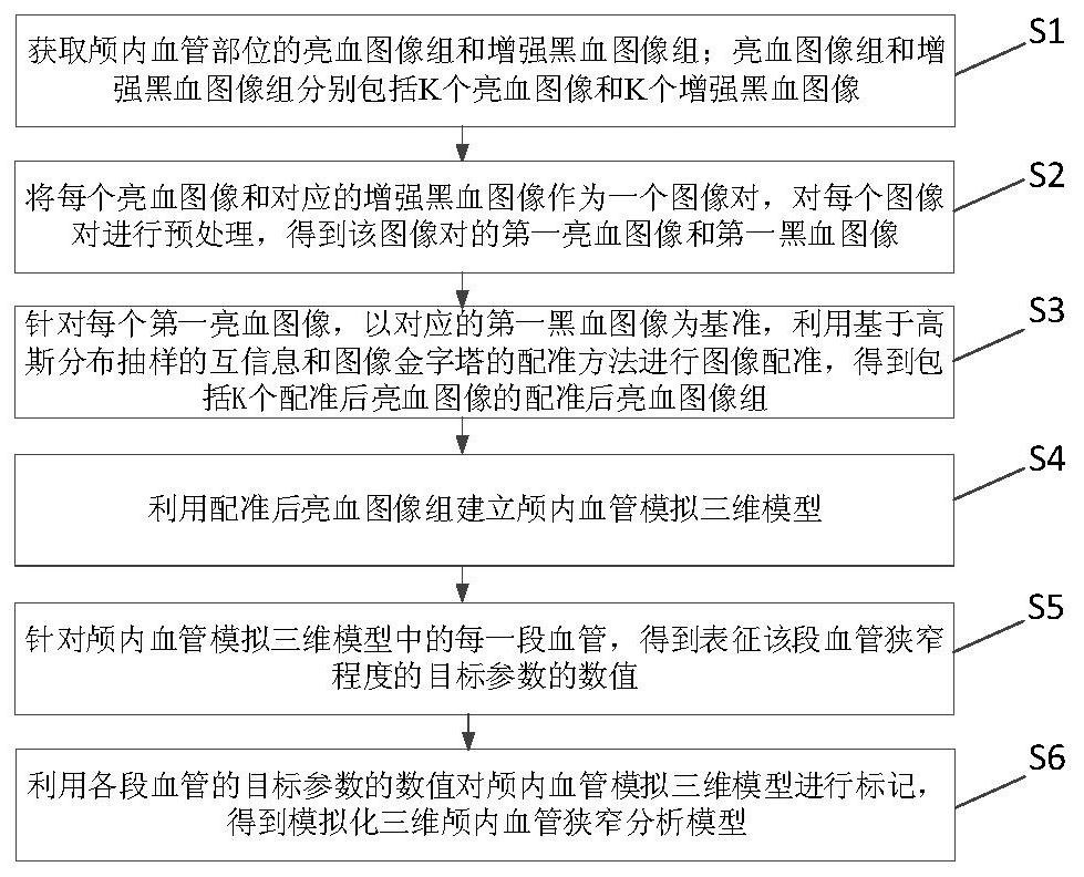
The invention discloses a method for establishing a simulated three-dimensional intracranial vascular stenosis analysis model. The method comprises the following steps: acquiring a bright blood imagegroup and an enhanced black blood image group of an intracranial vascular part; taking each bright blood image and the corresponding enhanced black blood image as an image pair, and preprocessing eachimage pair to obtain a first bright blood image and a first black blood image of the image pair; for each first bright blood image, taking the corresponding first black blood image as a reference, and performing image registration by using mutual information based on Gaussian distribution sampling and a registration method of an image pyramid to obtain a registered bright blood image group; establishing an intracranial blood vessel simulation three-dimensional model by using the registered bright blood image group; for each section of blood vessel in the intracranial blood vessel simulation three-dimensional model, obtaining a numerical value of a target parameter representing the stenosis degree of the section of blood vessel; and marking the intracranial vessel simulation three-dimensional model by utilizing the values of the target parameters of each section of vessel to obtain a simulated three-dimensional intracranial vessel stenosis analysis model.

Description

technical field [0001] The invention belongs to the field of image processing, and in particular relates to a method for establishing a simulated three-dimensional intracranial vascular stenosis analysis model. Background technique [0002] Medical data show that China is a high-incidence area of ​​intracranial artery stenosis in the world, and 33% to 50% of strokes and more than 50% of transient ischemic strokes (TIA) have symptomatic intracranial atherosclerotic stenosis (slCAS). ). [0003] At present, methods based on lumen imaging are usually used to evaluate the degree of intracranial vascular lesions and vascular stenosis clinically, such as digital subtraction angiography (Digital Subtraction Angiography, DSA), CT angiography (Computed Tomography Angiography, CTA). ), magnetic resonance angiography (Magnetic Resonance Angiography, MRA) and high-resolution magnetic resonance angiography (High-Resolution Magnetic Resonance Angiography, HRMRA), etc. The intracranial a...

Claims


Application Information

Patent Timeline
IPC IPC(8): G06T7/00G06T7/13G06T7/33G06T3/40G06T5/30G06T17/00G16H30/20G16H30/40
CPCG06T7/0012G06T7/13G06T7/344G06T3/4007G06T5/30G06T17/00G16H30/20G16H30/40G06T2207/10088G06T2207/20016G06T2207/20024G06T2207/30101
Inventor 贾艳楠王文杰
Owner XIAN CREATION KEJI CO LTD
Why Eureka
Try Eureka