High-resolution microscopic endoscope image cell nucleus segmentation method based on deep learning

A cell nucleus and endoscope technology, applied in the field of image processing, can solve the problem of low accuracy of cell nucleus segmentation, and achieve the effect of alleviating practical application requirements, improving accuracy, and improving the accuracy of cell nucleus segmentation

Active Publication Date: 2021-02-23
ZHEJIANG LAB +1
View PDF5 Cites 6 Cited by
  • Summary
  • Abstract
  • Description
  • Claims
  • Application Information

AI Technical Summary

Problems solved by technology

[0004] Aiming at the problems existing in the prior art, the present invention proposes a deep learning-based cell nucleus segmentation method for microendoscopic images, through a high-resolution network and a layered multi-scale attention mechanism, which integrates features of different levels to realize cell nucleus boundary segmentation Accurate segmentation, solving the problem of low accuracy of the prior art in microendoscopic image nucleus segmentation, and reducing the cost of manual processing

Method used


Image

  • High-resolution microscopic endoscope image cell nucleus segmentation method based on deep learning
  • High-resolution microscopic endoscope image cell nucleus segmentation method based on deep learning
  • High-resolution microscopic endoscope image cell nucleus segmentation method based on deep learning

Examples


Embodiment Construction

[0021] In order to make the object, technical solution and advantages of the present invention more clear and definite, the present invention will be further described in detail below with reference to the accompanying drawings. It should be understood that the specific embodiments described here are only used to explain the present invention, not to limit the present invention. The block diagrams shown in the drawings are merely functional entities and do not necessarily correspond to physically separate entities.

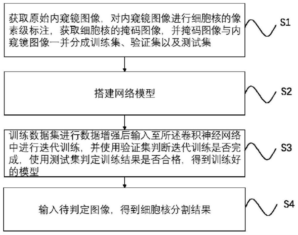
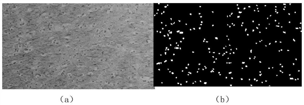
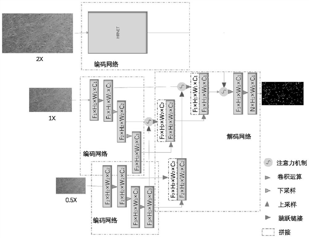
[0022] The present invention proposes a deep learning-based cell nucleus segmentation method for microendoscopic images to help users and their doctors quickly complete quantitative analysis of endoscopic images (such as cell or nucleus size, shape, density, quantity, and polymorphism) sex, etc.). Such as figure 1 The flow chart of the method for segmenting the nucleus of the microendoscope image specifically includes the following steps:

[0023] (1) Firstly, ...


PUM


Abstract

The invention discloses a high-resolution microscopic endoscope image cell nucleus segmentation method based on deep learning, and the method comprises the steps: obtaining an original endoscope image, carrying out the pixel-level marking of a cell nucleus of the endoscope image, and obtaining a mask image of the cell nucleus, dividing the marked mask image and the endoscope image into a trainingset and a verification set; constructing a hierarchical multi-scale attention mechanism high-resolution convolutional neural network model; inputting a training data set into the convolutional neuralnetwork for iterative training after data enhancement, and judging whether iterative training is completed by using a verification set; when it is judged that iterative training is completed, inputting the original endoscope image into the trained convolutional neural network, outputting the prediction probability that all pixels in the endoscope image belong to cell nucleuses, obtaining a segmentation result of the cell nucleuses, and realizing accurate segmentation of the input image.

Description

technical field [0001] The invention relates to the technical field of image processing, in particular to a deep learning-based cell nucleus segmentation method for microendoscopic images. Background technique [0002] An endoscope is an optical device that can detect the internal image information of an object. Medical endoscopes can enter the human body through the natural channels of the human body or small surgical incisions, and can help doctors see the internal structure of the human body that cannot be displayed by X-rays. It is an essential medical equipment for detection and diagnosis of lesions in the body and minimally invasive surgery, and is widely used in various fields of clinical medicine. Ordinary endoscopes observe internal tissues on a macro scale, identify suspicious areas, and if necessary, clamp suspicious tissues outside the body for histopathological diagnosis. This is a traumatic process, often accompanied by bleeding, infection, early missed diagnos...

Claims


Application Information

Patent Timeline
IPC IPC(8): G06T7/136G06N3/04G06N3/08G06T5/50
CPCG06T7/136G06T5/50G06N3/08G06T2207/10068G06T2207/30024G06T2207/20081G06T2207/20084G06T2207/20221G06N3/047G06N3/045
Inventor 王立强牛春阳杨青袁波张伟
Owner ZHEJIANG LAB
Why Eureka
Try Eureka