
Method for accurately and quantitatively detecting autophagy flux

A quantitative detection and autophagy technology, which is applied in botany equipment and methods, biochemical equipment and methods, viruses/bacteriophages, etc., can solve problems such as the inability to rule out systematic errors in reading values ​​between groups and limited application range, and achieve expansion Scope of application and detection accuracy, save time and cost, and promote the effect of research and development

Inactive Publication Date: 2021-02-05
SHENZHEN UNIV
View PDF3 Cites 2 Cited by
  • Summary
  • Abstract
  • Description
  • Claims
  • Application Information

AI Technical Summary

Problems solved by technology

[0010] The technical problem to be solved by the present invention is: how to provide a method for accurate and quantitative detection of autophagic flow, and solve the problem that the existing method cannot eliminate the systematic error caused by the reading value between groups, resulting in a severely limited application range

Method used


Image

  • Method for accurately and quantitatively detecting autophagy flux
  • Method for accurately and quantitatively detecting autophagy flux
  • Method for accurately and quantitatively detecting autophagy flux

Examples


Embodiment 1

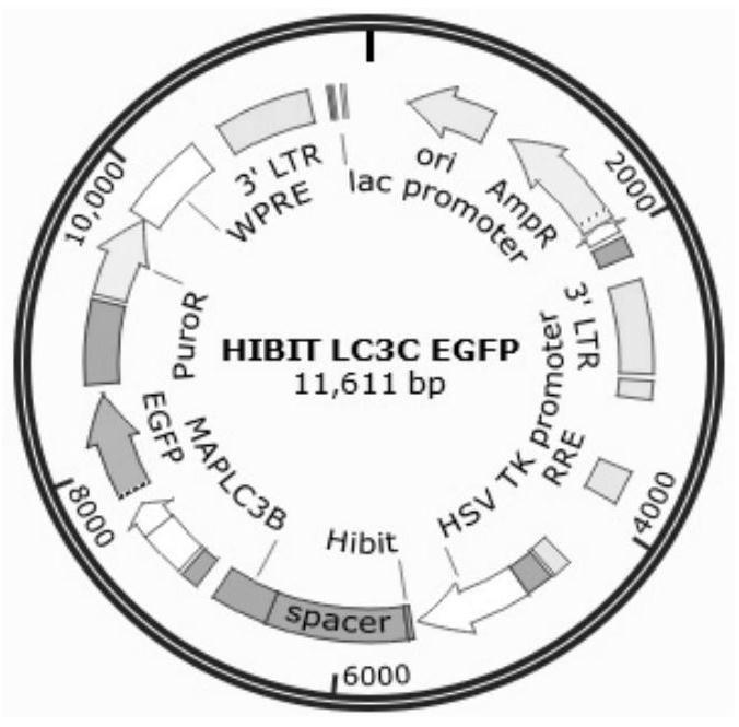
[0026] 1. Construct EGFP / LC3 HIBIT co-expression vector. pattern such as figure 2 shown.

[0027] Specific implementation steps: According to the method of seamless cloning, design independent EGFP fragments containing promoters and LC3HIBIT linearization primers respectively, and use KOD enzyme to amplify. System: 10×Buffer for KOD-Plus-5μL, 2mMdNTPs 5μL, 25mM MgSO4 2μL, Primer F 1.5μL, Primer R 1.5μL, Template DNA XμL (Plasmid DNA1-50ng / 50μL), KOD-Plus-1μL, water up to 50μL . Program: 94°C, 2min; 94°C, 15s, Tm-[5-10]°C, 30sec, 35cycle; 68°C, 1min. / kb. Electrophoresis and recovery of target fragments. Then prepare a seamless cloning reaction system: fragment molar ratio 1:1, 5×TEDA 4 μL, water up to 20 μL. Program: 30°C, 40min. Finally, the correct clones were screened by transformation sequencing.

[0028] 2. Transiently transfect or package lentivirus to infect cells, so that the cells co-express EGFP and LC3-HIBIT proteins. Specific implementation steps: transient...


PUM


Abstract

The invention discloses a method for accurately and quantitatively detecting an autophagy flux. The method comprises the following steps: (1) constructing an EGFP / LC3HIBIT co-expression vector, and respectively applying EGFP and LC3HIBIT to independent promoters without mutual influence; (2) obtaining co-expressed cells of EGFP and LC3-HIBIT proteins by a transient transfection or monoclonal screening method; (3) after screening the monoclonal stably transfected cells, carrying out various genetic operations related to autophagy on the basis of the cells, and screening autophagy related drugsand the like; and (4) setting programs on a multifunctional microplate reader to respectively measure luminescence and fluorescence readings, and calibrating a HIBIT reading caused by cell differencesby taking the EGFP as an internal reference in the experiment so as to accurately reflect the autophagy degree of the cells.

Description

technical field [0001] The invention belongs to the technical field of molecular biology, and in particular relates to a method for accurately and quantitatively detecting autophagy flow. Background technique [0002] Autophagy is an important process for the turnover of intracellular substances. Cells can eliminate, degrade and digest damaged, denatured, aged and non-functional cells, organelles and biomacromolecules such as denatured proteins and nucleic acids through autophagy and lysosomes. Provide the necessary raw materials for the reconstruction, regeneration and repair of cells, and realize the recycling and reuse of cells. It is both a "garbage processing plant" and a "waste recycling station" in the body; it can not only resist the invasion of pathogens, but also protect cells from damage by intracellular toxins. [0003] LC3B is a key gene in the process of autophagy. In the cell, LC3B is cleaved at the carboxy-terminus by Atg4, which has endoproteinase activit...

Claims


Application Information

Patent Timeline
Patent Type & Authority Applications(China)
IPC IPC(8): C12N15/867C12N15/65
CPCC12N15/86C12N15/65C12N2740/15043C12N2800/107
Inventor 张昊星李延鹏李嘉恒吴精博
Owner SHENZHEN UNIV
Learn More