
Detection device for pathological submission specimen

A technology of detection device and monitoring device, which is applied to measurement devices, collaborative operation devices, lubrication indicating devices, etc., can solve problems such as damage to fixative fluid, difficulty in monitoring the standardization of pathological specimen fixation and processing, etc., and achieve the effect of reducing operational risks.

Active Publication Date: 2021-01-29
PEOPLES HOSPITAL OF DEYANG CITY
View PDF7 Cites 1 Cited by
  • Summary
  • Abstract
  • Description
  • Claims
  • Application Information

AI Technical Summary

Problems solved by technology

[0003] The purpose of the present invention is to provide a detection device for pathological specimens sent for inspection, which solves the problem that the existing pathological specimens are difficult to monitor in the fixation process and the fixative is harmful to the human body.

Method used


Image

  • Detection device for pathological submission specimen
  • Detection device for pathological submission specimen
  • Detection device for pathological submission specimen

Examples


Embodiment 1

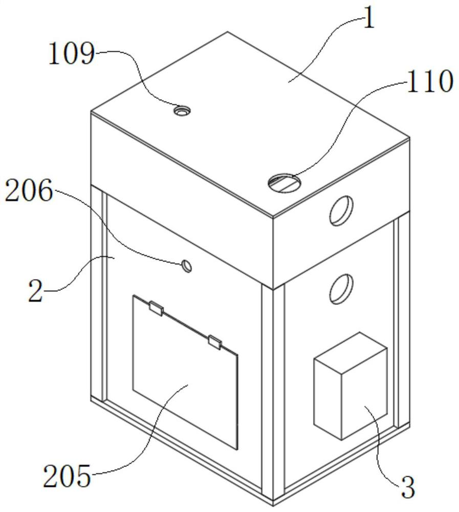
[0036] see figure 1 As shown, the present invention is a detection device for pathological inspection specimens, including a flushing device 1, a curing weighing device 2 and a monitoring device 3, the lower surface of the flushing device 1 is connected to the curing weighing device 2, and the monitoring device 3 is installed Only on the side of the curing and weighing device 2, the flushing device 1 performs flushing and pretreatment on the pathological specimens, and the pathological specimens after the flushing and pretreatment are added with curing liquid in the curing and weighing device 2, and the monitoring device 3 is used to record when the curing liquid is added. Manipulate and solidify environment data.

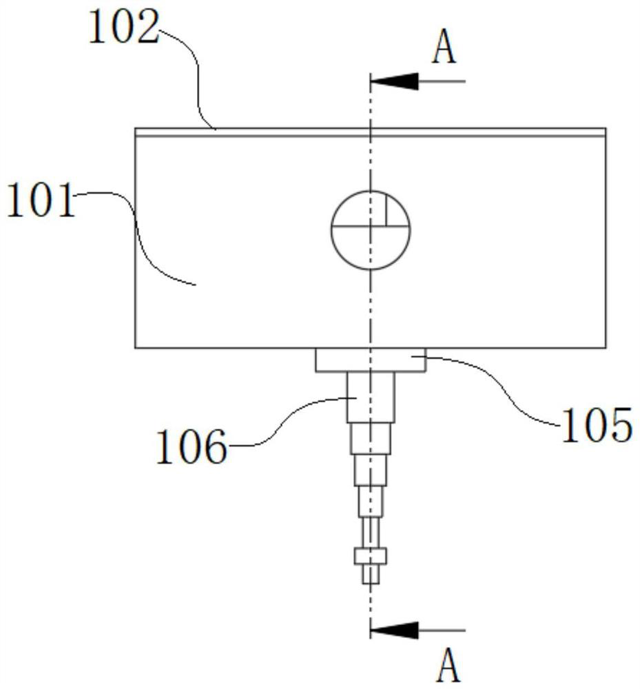
[0037] like Figure 2-6As shown, the flushing device 1 includes a fixed seat 101, a top cover 102, a connecting seat 105 and a telescopic rod 106. The fixed seat 101 cooperates with the top cover 102 to form a sealed box structure. The inner bottom surface of the ...

Embodiment 2

[0048] see Figure 1-9 As shown, the present invention is a detection device for pathological specimens, preferably, electronic scales, video cameras and infrared detectors are selected according to the quality and size of pathological specimens; its working principle and method of use are as follows: : first cut the pathological specimen into the required size, then place it on the material receiving plate 131 from the feed port 110, then cover the flushing cover 104 on the flushing seat 103, and then pump the flushing liquid to the flushing cap 104 by a suction pump Inside, rinse according to the time required for the pathological specimen to be rinsed, then turn off the suction pump, transfer the washed pathological specimen from the discharge port 107 to the pathological specimen container in the curing weighing device, and then adjust the height of the telescopic rod 106, Inject the fixative solution into the pathological sample container, and confirm the quality of the f...


PUM


Abstract

The invention discloses a detection device for a pathological submission specimen, and relates to the technical field of pathological specimen treatment. The detection device comprises a flushing device, a curing weighing device and a monitoring device, wherein the lower surface of the flushing device is connected with the curing weighing device, and the monitoring device is only installed on oneside face of the curing weighing device. The washing device is arranged to pretreat the pathological specimen, then the pretreated pathological specimen is transferred into the solidification weighingdevice to be injected with the stationary liquid, the monitoring device is arranged to monitor whether operation is standard or not when the stationary liquid is injected into the pathological specimen in real time, recording is conducted, and follow-up operation backtracking is facilitated; and the process of injecting the curing liquid is isolated through mutual cooperation of the flushing device and the curing weighing device, and the operation risk is reduced.

Description

technical field [0001] The invention belongs to the technical field of pathological specimen processing, in particular to a detection device for pathological examination specimens. Background technique [0002] In order to study the patient's condition, it is often necessary to remove the patient's pathological tissue and fix it within a specified period of time to prevent the cell tissue from being dissolved and the structure of the pathological tissue cannot be observed, so that the patient's condition cannot be accurately judged. Due to the different pathological tissues, different reagents are needed for fixation, and these reagents are often harmful to the human body; and because the curing reagents of different pathological tissues are different, the curing operation is also different, so when the curing of pathological specimens fails, it is difficult to identify On the other hand, it is not easy to trace back which step of the solidification operation is wrong, so a ...

Claims


Application Information

Patent Timeline
IPC IPC(8): G01G17/04G01F23/292G01J5/00G01N1/28G01N1/34A61L9/20G16H10/40G16H10/60G06K17/00
CPCG01G17/04G01F23/292G01J5/00G01N1/286G01N1/34A61L9/20G16H10/40G16H10/60G06K17/00G01N2001/2873
Inventor 乔明彪陈惠娟
Owner PEOPLES HOSPITAL OF DEYANG CITY
Learn More