
Posterior spinal reconstruction device used after laminectomy

A technology of resection and laminectomy, applied in the direction of spinal implants, prostheses, manufacturing tools, etc., can solve problems such as spinal instability, spinal canal restenosis, etc., and achieve a strong shaping effect

Pending Publication Date: 2021-01-01
FOURTH MILITARY MEDICAL UNIVERSITY
View PDF10 Cites 0 Cited by
  • Summary
  • Abstract
  • Description
  • Claims
  • Application Information

AI Technical Summary

Problems solved by technology

[0005] The purpose of the present invention is to provide a method that can solve the problem of restenosis of the spinal canal caused by spinal instability and scar adhesion after laminectomy. By filling the resection site with a 3D printed artificial lamina, on the one hand, it will produce a shielding effect and prevent surgery. The tissue behind the posterior spinal cord grows in; on the other hand, the degradation of the lamina occurs synchronously with the formation of bone, and finally completes the bony remodeling of the lamina

Method used


Image

  • Posterior spinal reconstruction device used after laminectomy

Examples


Embodiment Construction

[0018] The present invention will be described in further detail below in conjunction with the accompanying drawings.

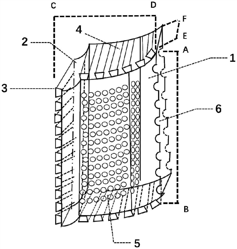
[0019] see figure 1 , the present invention is a 3D printing assembled degradable artificial lamina, the raw material used is polycaprolactone-tricalcium phosphate mixture (w / w 2:8-5:5), and the lamina degradation time is about 24 -36 months. The printing technology used is fusion extrusion molding (FDM), and the entire artificial lamina is designed in one piece. The main body 1 of the artificial lamina is in a semi-arc shape, and the parameters of the lamina vary according to different positions. Cervical lamina length (AB) is 10-15mm, transverse span (CD) is 10-15mm, lamina thickness (EF) is 3-5mm, arch height is 10-12mm; thoracic and lumbar lamina length is 15-30mm , the transverse span is 10-15mm, the thickness of the lamina is 6-8mm, and the arch height is 10-12mm. The main body of the lamina is divided into inner and outer layers, the inner layer is...


PUM

PropertyMeasurementUnit
pore sizeaaaaaaaaaa
lengthaaaaaaaaaa
thicknessaaaaaaaaaa

Abstract

The invention relates to a posterior spinal reconstruction device after laminectomy. The posterior spinal reconstruction device comprises a semi-arc vertebral plate body adopting 3D printing, whereinthe vertebral plate body is divided into two layers, the inner layer is a compact layer, the periphery of the outer layer is a compact layer, the center is a porous structure layer with the porosity of 50-80% and the pore diameter of 400-800 [mu]m, and the vertebral plate body adopting 3D printing adopts a mixture of polycaprolactone and tricalcium phosphate in the mass ratio being 2 to (8-5)to 5as the main raw material. The artificial vertebral plate is integrally designed through a 3D printing technology, and the formability is high. A porous structure is printed at the middle of the outerlayer of the vertebral plate, bone cells can crawl and grow into a stent after cancellous bone filling, and osseointegration and bone reconstruction of the stent are facilitated. Vertebral plate splicing teeth are printed on the edge of the vertebral plate and the joints of upper parts and lower parts and used for transverse and longitudinal assembly of the artificial vertebral plate. A fixing groove is printed in the outer side edge of the vertebral plate and can be buckled to a pedicle screw tail to complete fixing. The raw material adopted for vertebral plate printing is a completely-degradable polycaprolactone and tricalcium phosphate mixture, and the in-vivo complete degradation time is about 2-3 years.

Description

technical field [0001] The invention relates to a medical device, in particular to a device for reconstructing a posterior spinal path after laminectomy. Background technique [0002] Spinal stenosis is a disease in which the volume of the spinal canal decreases due to various reasons, thereby compressing the dural sac, spinal cord or nerve roots, resulting in a series of neurological symptoms such as pain, numbness, and weakness of the limbs. Decompressive laminectomy can effectively solve this clinical problem by removing the lamina, exposing the lamina and removing the lesion in the spinal canal. After the laminectomy, the structure of the posterior column of the spine is destroyed, resulting in instability of the spine. At the same time, the spinal cord and dural sac are exposed due to the loss of the protection of the laminae. In some patients, scar tissue forms behind the defect area, which grows into the spinal canal defect area, causing restenosis, and adhesion to t...

Claims


Application Information

Patent Timeline
Patent Type & Authority Applications(China)
IPC IPC(8): A61F2/44A61F2/28
CPCA61F2/44A61F2/28A61F2002/448A61F2002/30985A61F2002/30062A61F2002/2835
Inventor 郭征吴昊伍苏华
Owner FOURTH MILITARY MEDICAL UNIVERSITY
Learn More