
Fundus image processing system and method for cataract diagnosis

A fundus image and processing method technology, which is applied in image data processing, image enhancement, image analysis, etc., can solve problems such as low efficiency of manual inspection, and achieve the effects of reducing labor input costs, fast processing, and high real-time performance

Pending Publication Date: 2020-12-08
NING BO EYE HOSPITAL
View PDF9 Cites 0 Cited by
  • Summary
  • Abstract
  • Description
  • Claims
  • Application Information

AI Technical Summary

Benefits of technology

The technical effect described by this patented technology allows medical professionals to identify individuals with eye problems earlier than they are able or have difficulty detect them through visual examination alone. This helps prevent further complications such as vision loss from retinal detachment caused by glaucoma surgery.

Problems solved by technology

This patented technical problem addressed in this patents relates to finding effective ways to diagnose or treat cataractic conditions without requiring expensive equipment like an endoscope.

Method used


Image

  • Fundus image processing system and method for cataract diagnosis
  • Fundus image processing system and method for cataract diagnosis

Examples


Embodiment

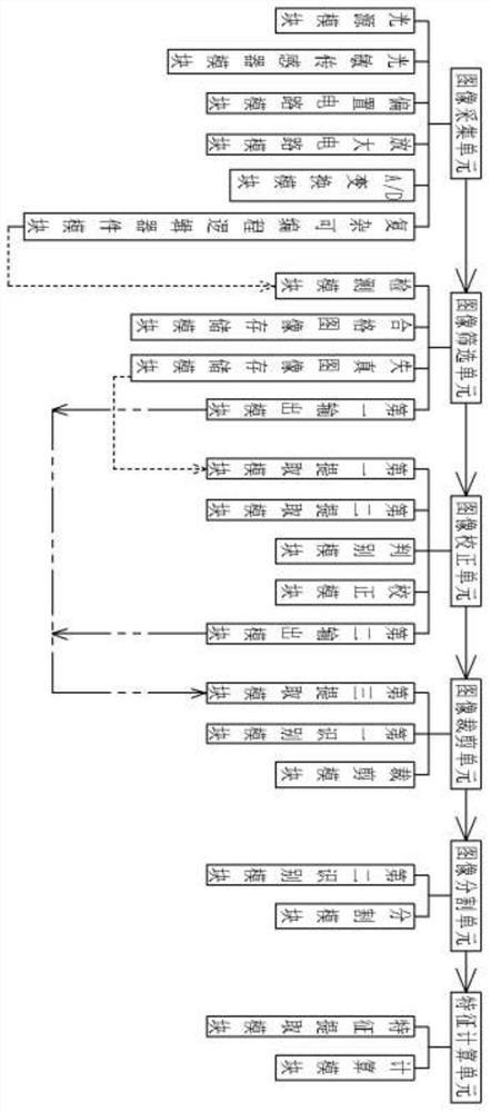
[0028] Example: such as figure 1 The shown fundus image processing system for cataract diagnosis includes an image acquisition unit for collecting image information of the patient's eyes, an image screening unit for screening the collected image information and marking distorted image information, An image correction unit for correcting the collected distorted image information, an image cropping unit for region cropping the fundus image corrected by the image correction unit, an image segmentation unit for further removing the clipping residual region, and A feature calculation unit for performing feature calculation on the final fundus image;

[0029] The image acquisition unit includes a light source module for light compensation during image acquisition, a photosensitive sensor module for sensing light on the patient's fundus, a bias circuit module, an amplification circuit module, and an amplifier circuit module for adjusting, amplifying, and converting the signal of the ...

experiment example

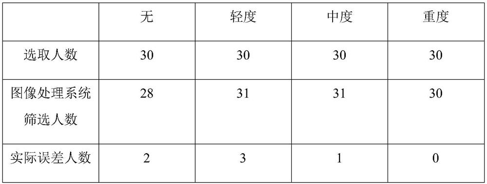
[0044] Experimental example: Cataract patients diagnosed with different degrees of illness and other ophthalmic disease patients without cataract symptoms were selected in an eye hospital in a city to conduct simulation processing experiments using the fundus image processing system of this embodiment. The specific processing results are as follows:

[0045]

[0046] In actual processing, the number of errors is specifically: 2 patients without cataracts are classified as mild patients, and 1 patient with mild cataracts is identified as moderate patients.

[0047] Conclusion: It can be seen from the above table that the total number of actual errors is 6 people, accounting for 5% of the total number of people, so the actual accuracy rate of this experiment is 95%.


PUM


Abstract

The invention discloses a fundus image processing system and method for cataract diagnosis. The processing system comprises an image acquisition unit, an image screening unit, an image correction unit, an image cutting unit, an image segmenting unit and a feature calculation unit. The method comprises the following steps: shooting fundus images by using the image acquisition unit; detecting the image information features by using the image screening unit, and dividing the fundus images into a distorted image and a qualified image; correcting the distorted image into a qualified image by usingthe image correction unit; cutting and segmenting the qualified fundus image by using the image cutting unit and the image segmenting unit; identifying the disease degree of the cataract by using thefeature calculation unit. The fundus image is screened after being processed, cataract patients can be screened out, the actual illness degree of the cataract patients can be determined, and popularization of ophthalmic examination in remote areas is facilitated.

Description


Claims


Application Information

Patent Timeline
Owner NING BO EYE HOSPITAL
Learn More