
Application of reduced graphene oxide film in frozen electron microscope

A technology of cryo-electron microscopy and graphene, which is applied in the direction of inorganic chemistry, carbon compounds, chemical instruments and methods, etc., can solve the problems of background noise, strong conductivity, etc., and achieve the effect of convenient acquisition, good resolution, and reduction of background noise

Pending Publication Date: 2020-11-10
TSINGHUA UNIV
View PDF10 Cites 0 Cited by
  • Summary
  • Abstract
  • Description
  • Claims
  • Application Information

AI Technical Summary

Problems solved by technology

[0006] Due to the super-strong electrical conductivity, mechanical properties, uniformity, and ultra-thin properties of graphene, it can partially or even completely eliminate the problems of strong background noise, poor electrical conductivity, and large molecules distributed at the gas-liquid interface in the current electron microscope grid.

Method used


Image

  • Application of reduced graphene oxide film in frozen electron microscope
  • Application of reduced graphene oxide film in frozen electron microscope
  • Application of reduced graphene oxide film in frozen electron microscope

Examples


Embodiment 1

[0035] Embodiment 1, the preparation of reduced graphene oxide electron microscopy grid support film

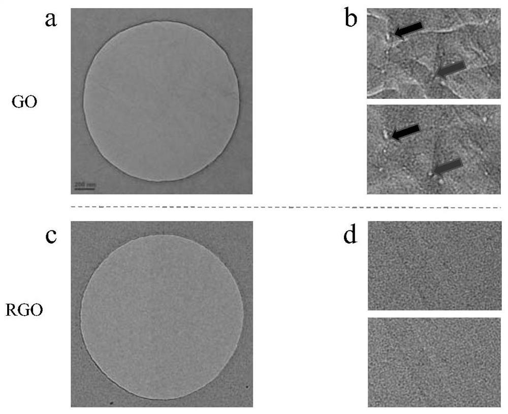
[0036] figure 1 (a) Schematic diagram of the preparation of the reduced graphene oxide grid for electron microscopy, the principle of which is to drop the solution containing graphene oxide into a container filled with water. The liquid level of the solution is higher than the grid placed in it, and the graphene oxide floats above the liquid level. Open the water outlet valve to make the liquid level drop slowly, and the graphene oxide is then attached to the carrier net. The graphene oxide electron microscope grid is naturally dried and then reduced at a high temperature at 300°C to prepare the reduced graphene oxide electron microscope grid. The specific process is as follows:

[0037] 1. Preparation of graphene oxide

[0038] Add H at a volume ratio of 9:1 to 3 g of graphene 2 SO 4 / H 3 PO 4 Mix the solution (400ml) and 18g potassium permanganate, heat to 50°C and k...

Embodiment 2

[0046] Embodiment 2, application of reduced graphene oxide electron microscope carrier network support film

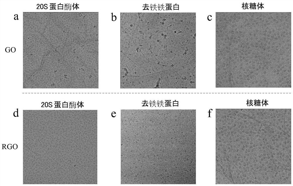
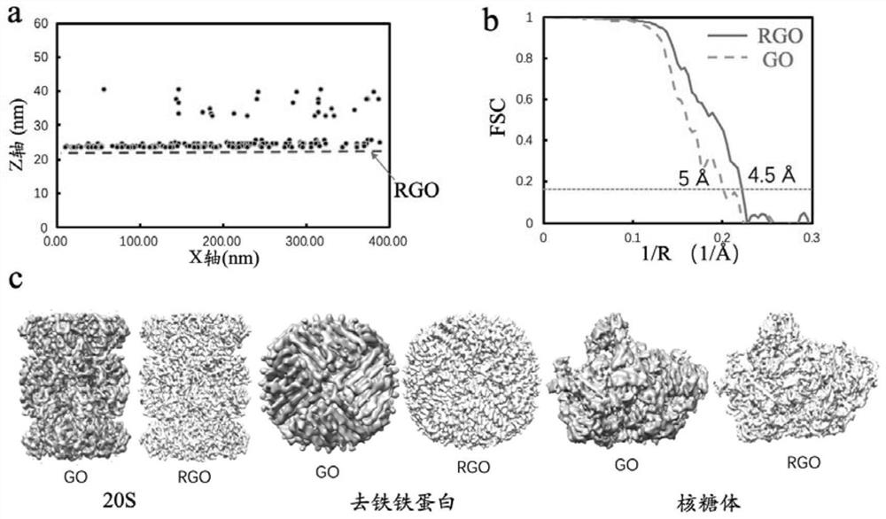
[0047] Take 20S proteasome, apoferritin and ribosome as examples to make frozen samples.

[0048] First, discharge and hydrophilize the grid covering the reduced graphene oxide prepared in Example 1: the vacuuming time during the treatment is 15s, and the low-grade discharge time is 15s. Then ~4ul 20S proteasome solution was added dropwise to the support membrane of the reduced graphene oxide electron microscope grid, and then transferred to FEI Vitrobot (frozen sample preparation instrument). The humidity of the Vitrobot cavity was adjusted to 100%, and the temperature was adjusted to 12°C. The suction time of the filter paper was set to 2s, and the force was set to -2. After the excess liquid was absorbed by the filter paper clip, the grid was quickly inserted into liquid ethane so that the water on the grid was quickly frozen to form glassy ice, and then transferr...


PUM


Abstract

The invention discloses an application of a reduced graphene oxide film in a frozen electron microscope. The reduced graphene oxide is used as or used for preparing a biological frozen electron microscope grid supporting film. The reduced graphene oxide is prepared according to a method comprising the following steps of transferring graphene oxide to an electron microscope carrying net, and reducing to obtain the reduced graphene oxide. Due to the defects of the existing supporting film, the preparation of a frozen sample is always a great challenge. Searching for a supporting film which is more stable, higher in conductivity and lower in background noise is the key point of work at the aspect of current sample manufacturing, and the reduced graphene oxide is the supporting film which mostaccords with the practical application at present, so that the defects of the conventional supporting film are overcome, and other characteristics such as ultrahigh mechanical strength and the like of reduced graphene oxide exist. It can be foreseen that the single-layer reduced graphene oxide carrying net can be produced at a large scale, and great convenience can be brought to sample preparation and later three-dimensional reconstruction of the frozen electron microscope.

Description

technical field [0001] The invention relates to the application of a reduced graphene oxide film in cryo-electron microscopy, and belongs to the technical field of biological cryo-electron microscopy. Background technique [0002] The long-term development of cryo-electron microscopy technology, especially the successive breakthroughs in data processing and camera hardware in recent years, has enabled this method to break through the limitations of previous resolutions, and structural analysis at near-atomic resolution has become more common. . This technique rapidly freezes a sample dissolved in water under physiological conditions, so that the sample is preserved in the amorphous glassy state of ice. Then observe with a transmission electron microscope and collect pictures. Finally, the three-dimensional reconstruction of molecules is carried out by combining relevant algorithms and image processing technology. Previously, this method was not widely used due to the inab...

Claims


Application Information

Patent Timeline
Patent Type & Authority Applications(China)
IPC IPC(8): G01N23/2005G01N23/20058G01N23/04G01N23/044C01B32/198
CPCG01N23/2005G01N23/20058G01N23/04G01N23/044C01B32/198G01N2223/03G01N2223/0565G01N2223/102
Inventor 王宏伟陈亚楠刘楠徐洁
Owner TSINGHUA UNIV
Learn More