
Method and device for identifying brain region position of fresh and live in-vitro tissue

An in vitro, fresh technology, applied in the field of neuroscience research experiments, can solve problems such as large errors, time-consuming and laborious, and researchers' troubles, so as to avoid differences between individuals, improve accurate identification, and improve accuracy.

Pending Publication Date: 2020-11-06
NANTONG UNIVERSITY
View PDF5 Cites 1 Cited by
  • Summary
  • Abstract
  • Description
  • Claims
  • Application Information

AI Technical Summary

Problems solved by technology

When using this type of brain atlas to identify and identify specific brain regions, it is often a great challenge for researchers. It usually requires experienced researchers to carefully compare the brain atlas with actual brain tissue slices in order to accurately identify the relevant brain regions. Area
In addition, due to differences in the age, weight, and gender of the experimental subjects used in the study, there are also certain deviations in the location of the brain regions. Fresh brain tissue is different from fixed and stained brain tissue sections. From the image, it is mainly gray and white. And it is usually 200-400 microns thick; and in actual operation, it is necessary to keep the neurons on the brain slices to maintain good activity, and it is necessary to perfuse artificial cerebrospinal fluid in real time. These factors allow the identification of specific brain regions on isolated fresh brain tissue slices It has brought great difficulties, which has caused serious troubles to researchers in the study of brain science, especially in the study of the physiological function of cells in specific brain regions in isolated fresh brain tissue slices
According to the traditional atlas recognition method, it is not only time-consuming and labor-intensive, but also often leads to large errors due to individual differences in animals and differences in the position of brain tissue slices.

Method used


Image

  • Method and device for identifying brain region position of fresh and live in-vitro tissue
  • Method and device for identifying brain region position of fresh and live in-vitro tissue

Examples


Embodiment Construction

[0031] In order to make the technical problems, technical solutions and advantages to be solved by the present invention clearer, the following will describe in detail with reference to the drawings and specific embodiments.

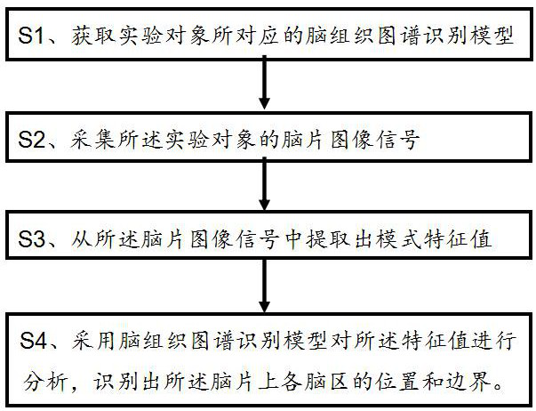
[0032] like figure 1 As shown, a method for identifying the brain region of a fresh isolated brain tissue comprises the following steps:

[0033] S1. According to the experimental subject from which the fresh isolated brain tissue comes from, obtain the brain tissue atlas recognition model corresponding to the experimental subject from the brain atlas model library.

[0034] S1-1. Pre-establishment of the brain atlas model library; the brain atlas model library includes brain tissue atlas recognition models corresponding to animals of different ages and genders;

[0035] S1-2. Acquiring the brain tissue atlas recognition model corresponding to the age group of the subject to which the fresh isolated brain tissue belongs.

[0036] Wherein, the pre-estab...


PUM


Abstract

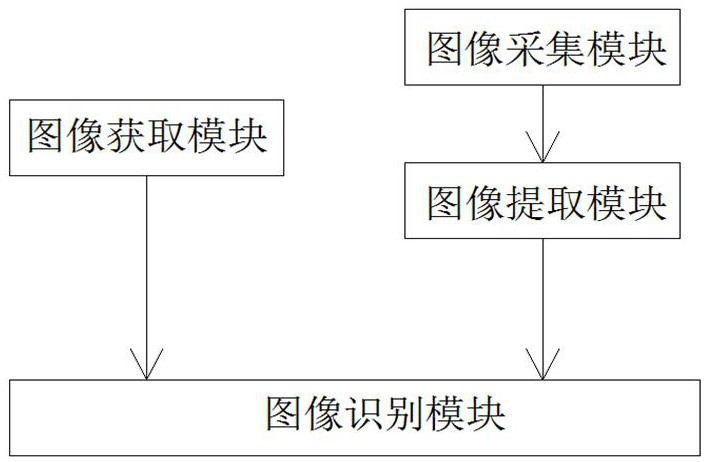
The invention discloses a method for identifying a brain region position of fresh and live in-vitro brain tissue. The method comprises the following steps: S1. obtaining a brain tissue atlas identification model corresponding to an experimental object from a brain atlas model library according to the experimental object where the fresh and live in-vitro brain tissue is derived; S2. acquiring brainslice image signals of the fresh and live in-vitro brain tissue; S3. extracting an eigen value of the brain slice image signals from the brain slice image signals; and S4. analyzing the extracted eigen value by adopting the brain tissue atlas identification model, so as to identify a brain atlas corresponding to a brain slice image of the fresh and live in-vitro brain tissue and identify variousbrain regions on the brain slice image. The invention discloses a device for identifying the brain region position of the fresh and live in-vitro brain tissue. The device comprises an image obtainingmodule, an image acquisition module, an image extraction module and an image identification module. According to the method and the device, the transformation to that a brain region structure of an in-vitro fresh and live brain tissue slice is observed, identified and labeled by machines from that the brain region structure of the in-vitro fresh and live brain tissue slice is observed and identified manually can be achieved.

Description

technical field [0001] The invention belongs to the technical field of neuroscience research experiments, and in particular relates to a method and a device for identifying brain regions of fresh and living isolated tissues. Background technique [0002] The brain is the most complex organ in animals. It is composed of a large number of neurons and other cells with different shapes and functions. Learning and memory, emotion, cognition, consciousness and other activities are encoded through different neurons and neural networks. Studying and understanding the brain is the basis for treating neuropsychiatric diseases, simulating brain functions, and optimizing artificial intelligence. With the emergence and mature application of immunohistochemical staining, fluorescent transgenic technology and other chromogenic methods, people have a more detailed understanding of the brain region structure of various model animals such as mice and rats, and have completed the mapping of th...

Claims


Application Information

Patent Timeline
IPC IPC(8): A61B5/00
CPCA61B5/0042A61B5/0073A61B5/4064
Inventor 吴小波高永静张志军姜保春李炜
Owner NANTONG UNIVERSITY
Learn More