Cerebral hemorrhage automatic detection method based on brain auxiliary image and electronic medium

An auxiliary image, automatic detection technology, applied in the field of image processing, can solve problems such as easy to cause errors, and achieve the effect of improving diagnostic efficiency, reducing technical requirements, and improving convenience

Active Publication Date: 2020-08-14
NANJING ANKE MEDICAL TECH CO LTD
View PDF5 Cites 5 Cited by
  • Summary
  • Abstract
  • Description
  • Claims
  • Application Information

AI Technical Summary

Problems solved by technology

However, in fact, there are many similarities between each type of cerebral hemorrhage. For example, the CT value is not much different, and there are many similarities in shape. Just relying on input cerebral hemorrhage images for training does not necessarily get very accurate r

Method used


Image

  • Cerebral hemorrhage automatic detection method based on brain auxiliary image and electronic medium
  • Cerebral hemorrhage automatic detection method based on brain auxiliary image and electronic medium
  • Cerebral hemorrhage automatic detection method based on brain auxiliary image and electronic medium

Examples


Embodiment Construction

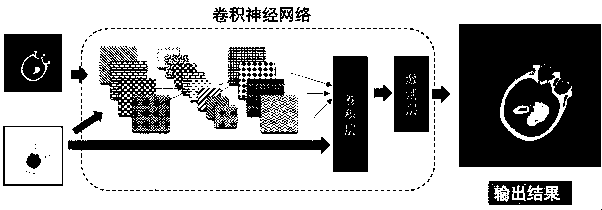
[0045] A method for automatic detection of cerebral hemorrhage based on brain auxiliary images and electronic media of the present invention will be further explained and described below in conjunction with the accompanying drawings.

[0046] as attached Figure 5 As shown, the existing cerebral hemorrhage includes the following types of hemorrhage, such as epidural hemorrhage, subdural hemorrhage, cerebral parenchymal hemorrhage, brainstem hemorrhage and subarachnoid hemorrhage and so on. In the patient's brain CT image, the pixel value of the normal area is 20-40HU, the pixel value of the epidural hemorrhage, subdural hemorrhage, cerebral parenchymal hemorrhage and brainstem hemorrhage is 45-75HU, and the subarachnoid hemorrhage The pixel value of the area is 40-75HU, so it is difficult to judge the type of bleeding based on the pixel value alone. Usually manual diagnosis is based on the pixel value, combined with the location of the bleeding area, and the shape of the blee...


PUM


Abstract

The invention discloses a cerebral hemorrhage automatic detection method based on a brain auxiliary image and an electronic medium, and is applied to the technical field of image processing, and the method comprises the steps: carrying out the CT scanning of the head of a patient, and obtaining a brain CT image; generating a brain auxiliary image according to the brain CT image; and inputting thebrain CT image and the brain auxiliary image into a pre-trained neural network, automatically detecting a cerebral hemorrhage area in the brain CT image in combination with the brain auxiliary image,and outputting a mask including the cerebral hemorrhage area and type. According to the invention, the brain auxiliary image and the brain CT image scanned by the patient are combined for use, the cerebral hemorrhage area and type identification accuracy of the neural network is improved, the technical requirements of doctors in the diagnosis process are reduced, and the diagnosis efficiency is greatly improved.

Description

technical field [0001] The invention relates to the technical field of image processing, in particular to an automatic detection method of cerebral hemorrhage based on an auxiliary brain image and an electronic medium. Background technique [0002] Computer-aided detection and analysis of cerebral hemorrhage in CT images is to identify and analyze the type and area of ​​cerebral hemorrhage in the image. It is very important for the follow-up treatment of stroke. The image processing software of cerebral hemorrhage in the prior art can detect the cerebral hemorrhage image and output the type and area of ​​cerebral hemorrhage, and the detection is mostly based on the method of artificial intelligence, that is, first construct the neural network, and then use the artificially marked data Set, and then use this data set to train the neural network. [0003] Currently public AI-assisted analysis methods for cerebral hemorrhage usually input the image of cerebral hemorrhage into...

Claims


Application Information

Patent Timeline
IPC IPC(8): G06T7/00
CPCG06T7/0014G06T2207/10081G06T2207/20081G06T2207/20084G06T2207/30016
Inventor 曾凯
Owner NANJING ANKE MEDICAL TECH CO LTD
Why Eureka
Try Eureka