
Method for separating and extracting amniotic membrane stem cells from placentas

A technology for stem cells and amniotic membrane, which is applied in the field of separating and extracting amniotic membrane stem cells from placenta, can solve the problems of increased production cost of amniotic membrane stem cells, low extraction rate of neural stem cells, complicated separation methods, etc., and achieves simple and fast processing steps and realizes waste utilization. , the effect of prolonging the acquisition time

Pending Publication Date: 2020-08-11
成都容医汇生物技术研究院
View PDF3 Cites 0 Cited by
  • Summary
  • Abstract
  • Description
  • Claims
  • Application Information

AI Technical Summary

Problems solved by technology

[0004] However, the acquisition of neural stem cells currently has problems such as low extraction rate, complicated separation methods, and scarce sources, which lead to increased production costs of amniotic stem cells

Method used


Image

  • Method for separating and extracting amniotic membrane stem cells from placentas
  • Method for separating and extracting amniotic membrane stem cells from placentas
  • Method for separating and extracting amniotic membrane stem cells from placentas

Examples


Embodiment 1

[0040] A method for separating and extracting amniotic stem cells from placenta, specifically comprising the following steps:

[0041] Step 1: Take out the placenta with sterile tweezers and place it on a sterile culture plate, wash the blood stains on the surface of the placenta with sterile distilled water containing penicillin and streptomycin, then rinse the placenta again with 0.9% normal saline, and then Cut a piece of amniotic membrane from the placenta after rinsing and transfer it to a new sterile plastic petri dish, and continue to wash the blood stains on the surface of the amniotic membrane with normal saline, in which penicillin and streptomycin The concentration is 100 units / mL.

[0042]Step 2: Cut the amniotic membrane washed in step 1 into 0.5-1cm 2 The small pieces of amniotic membrane were washed with normal saline, and then the washed small piece of amniotic membrane was transferred to a new sterile plastic petri dish and rinsed with normal saline until the...

Embodiment 2

[0056] Amniotic membrane stem cells were separated and extracted from the placenta according to the method of Example 1, the difference was that the digestion time in Step 5 and Step 9 was both 30 s, and the centrifuge in Step 11 was pre-cooled to 2°C.

Embodiment 3

[0058] The amniotic membrane stem cells were separated and extracted from the placenta according to the method of Example 1, the difference was that the digestion time in Step 5 and Step 9 was both 90 s, and the centrifuge in Step 11 was pre-cooled to 8°C.

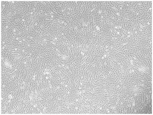
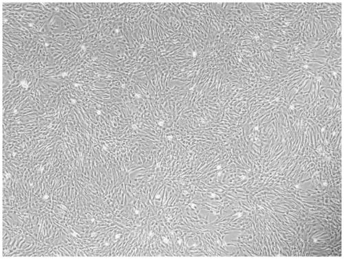
[0059] According to the above-mentioned Example 1 about a method for isolating and extracting amniotic membrane stem cells from placenta, under the same conditions, the present invention randomly selects nine full-term placentas of 25-30-year-old puerperas to isolate and extract amniotic membrane stem cells, and The obtained amniotic membrane stem cells are collected with the Olympus CX41+DP20 digital camera for amniotic membrane stem cell data collection. The amniotic membrane stem cell data collected are as follows: Figure 1-9 shown.

[0060] From Figure 1-9 It can be seen that the amniotic membrane stem cells isolated from 9 different placentas are all in the shape of typical stem cells, which meet the inspection stand...


PUM


Abstract

The invention discloses a method for separating and extracting amniotic membrane stem cells from placentas. The method is used for extracting the amniotic membrane stem cells by simply cleaning and sterilizing the waste placentas in hospitals, so that resources are fully utilized. The method comprises the following steps of firstly, cleaning, dicing, culturing and digesting amniotic membranes to obtain a small amount of the amniotic membrane stem cells from the amniotic membranes, and then continuously culturing, digesting and centrifuging the obtained amniotic membrane stem cells to obtain alarge amount of the amniotic membrane stem cells. The preparation method is simple, convenient and easy to operate; and when the amniotic membrane stem cells are extracted from the amniotic membranes,the amniotic membranes are sheared into meat paste of 2-3 mm<3>, and more amniotic membrane stem cells are promoted to be exposed, so that the contact area between the amniotic membrane stem cells and a culture medium is increased, in-vitro amplification of the amniotic membrane stem cells is promoted, and the amount of the amniotic membrane stem cells extracted from the amniotic membranes is increased.

Description

technical field [0001] The invention relates to a novel cell separation and purification method, in particular to a method for separating and extracting amniotic membrane stem cells from placenta. Background technique [0002] Amniotic stem cells have neurobiological functions and have homology with neural stem cells. Although neural stem cell (NSC) research started late, it is a hot spot in current research. The application of NSC in the treatment of nervous system diseases is limited due to the small number of NSC in adult nervous tissue, scattered distribution, difficulty in obtaining materials, difficulty in establishing non-immunogenic NSC cell lines, and immune rejection in allogeneic transplantation. With the gradual deepening of research on amniotic membrane epithelial cells having the characteristics of neural stem cells, amniotic membrane stem cells will bring new opportunities for the treatment of neurodegenerative diseases and traumatic nerve injuries. [0003]...

Claims


Application Information

Patent Timeline
IPC IPC(8): C12N5/073C12N5/0775
CPCC12N5/0605C12N5/0668C12N2509/00
Inventor 于坤刘鹏刘蕊李馨何前希王俊
Owner 成都容医汇生物技术研究院
Learn More