
Endoscope-type guide device for tracheal intubation

An endoscopic and endotracheal intubation technology, applied in the field of medical devices, can solve the problems of patient injury, high cost, complex structure, etc., and achieve the effects of ensuring stable observation, high safety usability, and convenient application

Pending Publication Date: 2020-07-17
THE FIRST AFFILIATED HOSPITAL OF MEDICAL COLLEGE OF XIAN JIAOTONG UNIV
View PDF4 Cites 2 Cited by
  • Summary
  • Abstract
  • Description
  • Claims
  • Application Information

AI Technical Summary

Problems solved by technology

[0002] Endotracheal intubation is a common clinical operation. The core operation step for successful intubation is to send the endotracheal tube into the trachea through the glottic fissure. Therefore, it is very important to identify and see the glottic fissure during the operation. The traditional operation method It is very inconvenient for the operator to hold the laryngoscope in one hand and raise the lower jaw to identify the glottic cleft with the naked eye under the weak light of the laryngoscope. Especially for some patients with short jaws, the glottic cleft may not be visible at all, so blind insertion is required. To confirm the position of the glottic cleft, the tracheal intubation tool in the existing design is equipped with a camera at one end. Although the inside of the oral cavity can be seen during the intubation process, the structure is very complicated, the cost is high, and it is not easy to use; The camera is very easy to shake, which is not conducive to the observation of medical staff; and the camera has no shell protection, which is easy to cause harm to the patient, and to a certain extent, reduces the operational efficiency of the endotracheal intubation process

Method used


Image

  • Endoscope-type guide device for tracheal intubation
  • Endoscope-type guide device for tracheal intubation
  • Endoscope-type guide device for tracheal intubation

Examples


Embodiment

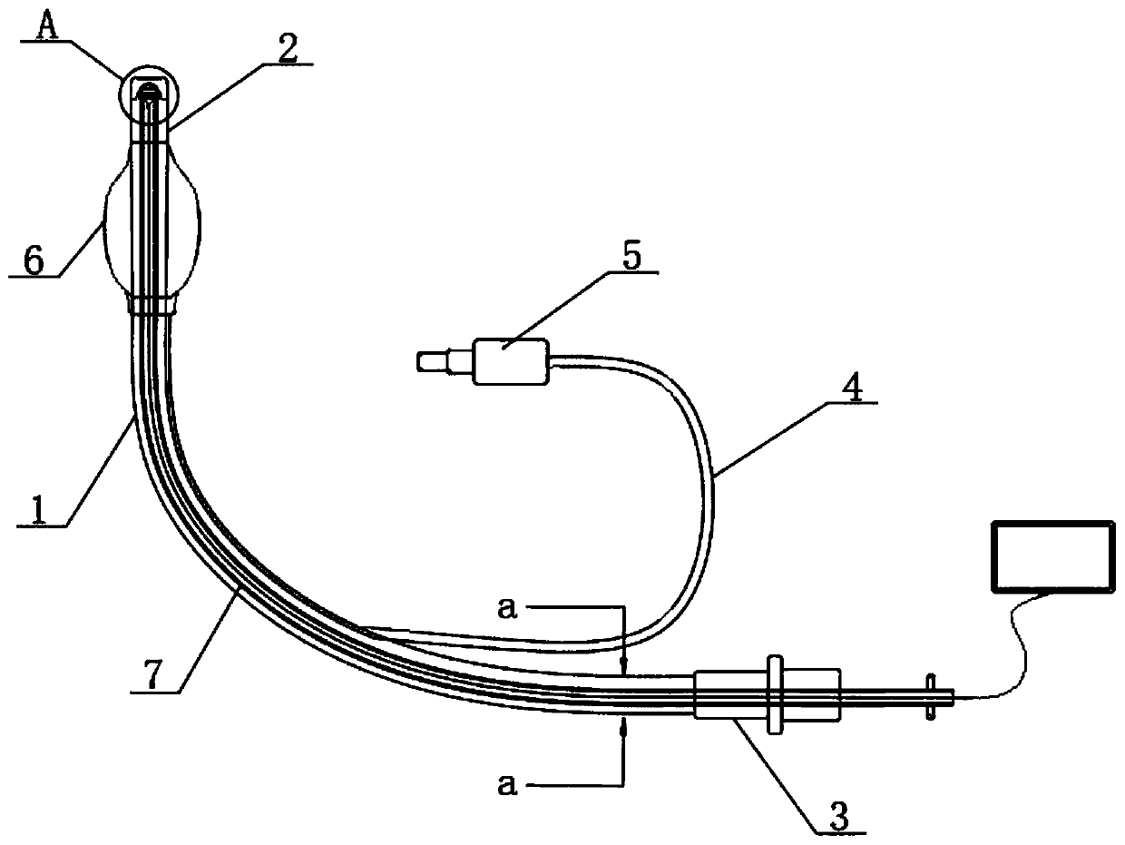
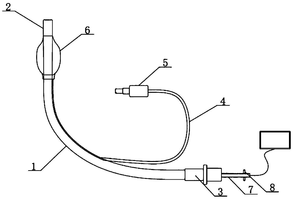
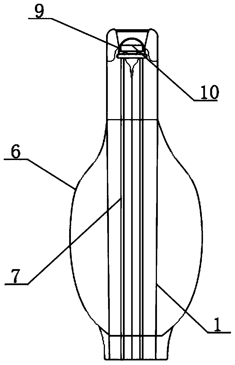
[0022] See Figure 1-6 The present invention provides the following technical solutions: an endoscopic guiding device for tracheal intubation, comprising a tube body 1, a joint 3, an inflation valve 5, and a balloon 6. The inflation valve 5 is installed along the inner wall of the tube body 1 through the catheter 4 Connected to the airbag 6, the tube body 1 is integrally formed with an inserting head 2 at one end different from the connector 3. A tube core 7 is installed inside the tube body 1, and a connecting block 17 is welded on both sides of the tube core 7 and a connecting block 17 The inside of the tube body 1 is connected with a thread 16 and the side of the tube core 7 is integrally formed with a clamping strip 14. The inside of the tube body 1 is provided with a matching slot 15 at a position corresponding to the clamping strip 14, and the tube core 7 The end part of the inserting head 2 is provided with a rotating cap 9, and the inside of the rotating cap 9 is equipp...


PUM


Abstract

The invention belongs to the technical field of medical instruments, and in particular to an endoscope-type guide device for tracheal intubation. The endoscope-type guide device comprises a tube body,a joint, an inflation valve and an air bag; the inflation valve is arranged along the inner wall of the tube body through a catheter and is connected with the air bag; an insertion head is integrallyformed at one end, which is different from the joint, of the tube body; a tube core is arranged in the tube body; and two sides of the tube core are respectively welded with connecting blocks. A wireless miniature camera at the end part of the tube core can quickly carry out image feedback through the penetrating connection effect of the tube core in the tube body, and medical personnel can conveniently use and operate the device after receiving image signals by using peripheral equipment. The device has a simple structure and is convenient to apply. Through utilization of the clamping connection effect of a clamping strip and clamping groove part and the clamping connection property of a clamping hole in an inner groove, the wireless miniature camera does not shake easily in an observation process, so that stable observation of medical personnel is ensured; and meanwhile, a certain external protection effect is given to the wireless miniature camera by external arrangement of a rotary cap.

Description

Technical field [0001] The invention belongs to the technical field of medical devices, and specifically relates to an endoscopic guiding device for tracheal intubation. Background technique [0002] Tracheal intubation is a common clinical operation. The core operation step for successful intubation is to send the tracheal intubation through the glottis into the trachea. Therefore, it is very important to identify and see the glottis during the operation. The traditional operation method It is inconvenient for the surgeon to hold the laryngoscope and lift the mandible to identify the glottis under the weak light of the laryngoscope. It is very inconvenient, especially for some patients with short mandibular, who may not be able to see the glottis at all, so blind insertion is required. Confirm the position of the glottis. The existing design of the tracheal intubation tool has a camera at one end. Although the inside of the oral cavity can be seen during the intubation process, ...

Claims


Application Information

Patent Timeline
IPC IPC(8): A61M16/04A61B1/267A61B1/05A61B1/06
CPCA61M16/0488A61B1/267A61B1/05A61B1/0661
Inventor 苗常青吴梦茹梁欢樊丹丹屈慧
Owner THE FIRST AFFILIATED HOSPITAL OF MEDICAL COLLEGE OF XIAN JIAOTONG UNIV
Learn More