
Full-automatic high-performance leukocyte segmentation method

A white blood cell, high-performance technology, applied in the field of image processing, can solve problems such as difficult segmentation of cytoplasmic regions, and achieve high recognition, easy recognition, and good performance

Pending Publication Date: 2020-06-19
张云超
View PDF2 Cites 3 Cited by
  • Summary
  • Abstract
  • Description
  • Claims
  • Application Information

AI Technical Summary

Problems solved by technology

[0011] 3.) The cytoplasmic region is very similar to the background, so it is difficult to segment the cytoplasmic region correctly

Method used


Image

  • Full-automatic high-performance leukocyte segmentation method
  • Full-automatic high-performance leukocyte segmentation method
  • Full-automatic high-performance leukocyte segmentation method

Examples


Embodiment Construction

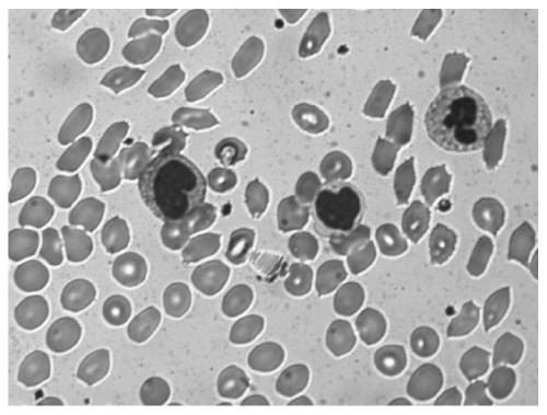
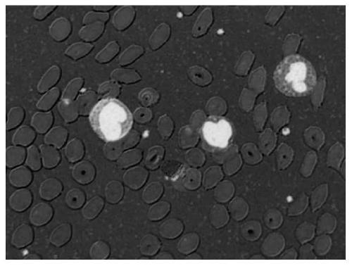
[0051] refer to figure 1 , in order to improve the accuracy and adaptability of the white blood cell segmentation algorithm, and solve or partially solve the above-mentioned problems, the present invention first collects the digital image of the blood smear stained by Wright, and amplifies it by 500 times, and then uses the following process Figure breakdown of white blood cells:

[0052] Step S100, converting the RGB image into an image in HSL (Hue, Saturation, Lightness) color space. The model of the HSL color space corresponds to a double cone and a sphere in the cylindrical coordinate system (white is at the upper vertex, black is at the lower vertex, and the center of the largest cross-section is half gray), and the image of the HSL color space includes hue, saturation , the brightness of three sub-images;

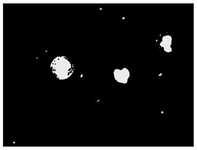
[0053] Step S200, detecting cell nuclei from the scaled saturation image;

[0054] Step S300, marking the cell nucleus area on the full-resolution sub-image;

[0...


PUM


Abstract

The invention provides a full-automatic high-performance leukocyte segmentation method. In clinical diagnosis and treatment of a patient, classification and counting of white blood cells on a blood smear are very important. However, manual leukocyte classification and counting by hematologists are time-consuming and error-prone. According to the algorithm provided by the invention, a series of processing such as segmentation is carried out on the nuclear region through the zoomed saturation image, and then image processing such as difference is carried out on the green image and the blue imagenormalized by the red image, so that the purpose of automatically and accurately segmenting the cytoplasm region in the sub-image is achieved.

Description

technical field [0001] The invention relates to the field of image processing, in particular to a fully automatic high-performance white blood cell segmentation method. Background technique [0002] White blood cells are an important part of the human immune system, and changes in the shape and number of white blood cells are usually used as a reference for diagnosis in clinical practice. [0003] In the past, most experienced hematology staff used optical microscopes to manually identify and classify white blood cells, and then judge the types of white blood cells based on information such as the nucleus, cytoplasm, and chromosome color in the microscopic white blood cells, and record the number of white blood cell types. . According to hospital statistics, about 20% of routine blood samples require manual microscopic review. This manual method takes a long time to adjust the focus and observe and identify each time, and the staff of the hematology department responsible ...

Claims


Application Information

Patent Timeline
Patent Type & Authority Applications(China)
IPC IPC(8): G06T7/187G06T7/90G06T7/62G06T5/30G06T5/40G06T7/00
CPCG06T7/187G06T7/90G06T7/62G06T5/30G06T5/40G06T7/0012G06T2207/10061
Inventor 张云超邱天
Owner 张云超
Learn More