Target area identification and necrotic tissue evaluation method and system, equipment and medium
A target area and tissue technology, applied in the computer field, can solve the problems of the thermal ablation process cannot be monitored in real time, cannot be popularized, and the imaging effect is poor, and achieves the effect of fast image acquisition, good presentation effect, and improved recognition accuracy.
- Summary
- Abstract
- Description
- Claims
- Application Information
AI Technical Summary
Problems solved by technology
Method used
Image

Examples
Embodiment 1
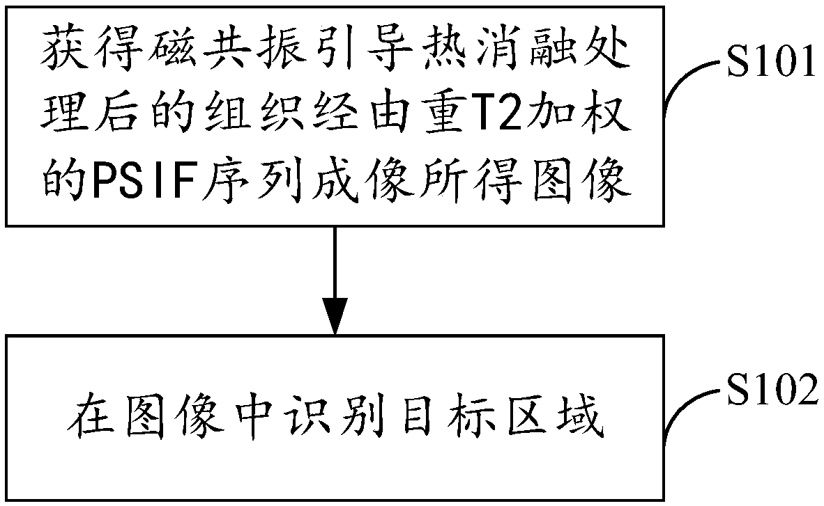
[0034] figure 1 It shows the implementation flow of the target area recognition method in the tissue imaging image provided by Embodiment 1 of the present invention. For the convenience of description, only the parts related to the embodiment of the present invention are shown, and the details are as follows:
[0035] In step S101 , an image obtained through a T2-weighted time-reversal steady-state free precession (Reserved Fast Imaging with Steady-state free Precession, PSIF) sequence imaging of the tissue after magnetic resonance-guided thermal ablation treatment is obtained.
[0036] In this embodiment, for the convenience of image analysis, it is hoped that the gray scale of the image is mainly determined by a specific imaging parameter, that is, weighted imaging will be used. In this embodiment, the gray scale of the image is mainly determined by the imaging parameter T2, that is, the T2-weighted imaging to highlight differences in tissue T2 transverse relaxation. T2 is ...
Embodiment 2
[0047] On the basis of Embodiment 1, this embodiment further provides the following content:
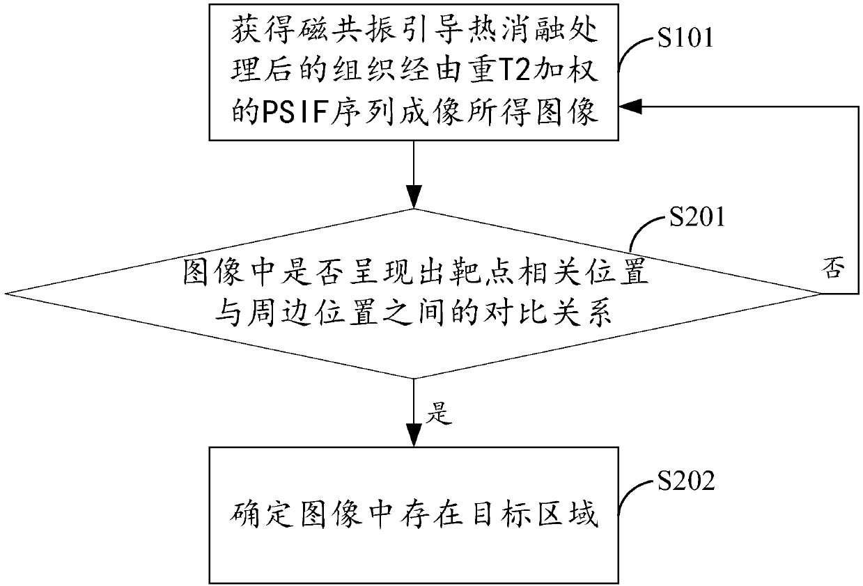
[0048] Such as figure 2 As shown, step S102 specifically includes the following steps:
[0049] In step S201, it is judged whether there is a contrast relationship between the relative position of the target point and the surrounding position in the image, and if so, execute step S202, otherwise, end the process or return to execute step S101.
[0050] In step S202, it is determined that there is a target area in the image.
[0051] In this embodiment, by scanning the grayscale within a specified range in the image, if it is detected that there is an obvious grayscale change from a certain position to another position, it is judged that the above-mentioned contrast relationship exists, and then it is determined that there is a target area in the image . The detection of obvious grayscale changes can introduce corresponding empirical thresholds, for example, if the grayscale diffe...
Embodiment 3
[0053] On the basis of Embodiment 2, this embodiment further provides the following content:
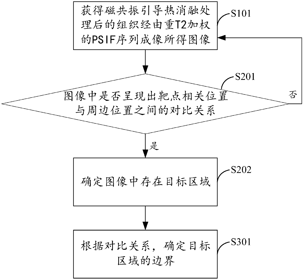
[0054] Such as image 3 As shown, step S102 specifically also includes the following steps:
[0055] In step S301, when it is judged in step S201 that the above contrast relationship exists, the boundary of the target area is determined according to the contrast relationship.
[0056] In this embodiment, the boundary of the target area can be determined and outlined at the position where obvious grayscale changes occur.
PUM
 Abstract
Description
Claims
Application Information
 - R&D
- Intellectual Property
- Life Sciences
- Materials
- Tech Scout
- Unparalleled Data Quality
- Higher Quality Content
- 60% Fewer Hallucinations
Browse by: Latest US Patents, China's latest patents, Technical Efficacy Thesaurus, Application Domain, Technology Topic, Popular Technical Reports.
© 2025 PatSnap. All rights reserved.Legal|Privacy policy|Modern Slavery Act Transparency Statement|Sitemap|About US| Contact US: help@patsnap.com