
Radiographic imaging apparatus

A photographing device and radiation technology, applied in the directions of clinical application of radiological diagnosis, instruments for radiological diagnosis, image enhancement, etc., can solve the problem that the progress status cannot be easily confirmed, the treatment target part that is difficult to confirm the treatment, and the treatment target part is not indicated. problems with marks

Pending Publication Date: 2020-05-19
SHIMADZU SEISAKUSHO CO LTD
View PDF8 Cites 0 Cited by
  • Summary
  • Abstract
  • Description
  • Claims
  • Application Information

AI Technical Summary

Problems solved by technology

[0005] However, in the X-ray diagnostic apparatus of the above-mentioned Patent No. 5337525, there is the following problem: when the blood vessel of the treatment target part is treated, there is no mark indicating the treatment target part, so it is difficult to confirm the blood vessel in the subject after the treatment. The treatment target site (dilated site of blood vessel) treated with the balloon
Therefore, there is a problem that it is not possible to easily confirm the progress of the treatment in the subject by dilation of the balloon.

Method used


Image

  • Radiographic imaging apparatus
  • Radiographic imaging apparatus
  • Radiographic imaging apparatus

Examples


Embodiment Construction

[0040] Embodiments of the present invention will be described below based on the drawings.

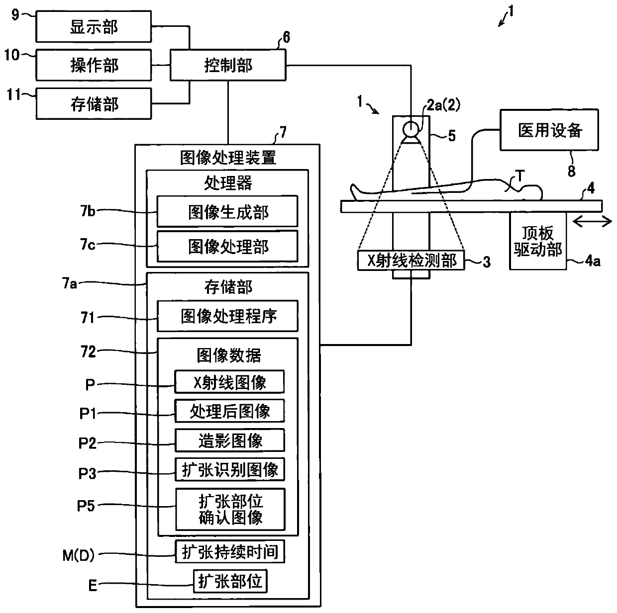
[0041] refer to Figure 1 ~ Figure 1 3, the configuration of the X-ray imaging apparatus 1 according to this embodiment will be described. The X-ray imaging apparatus 1 captures an X-ray image P (fluoroscopic image) of the inside of the subject T by irradiating the subject T such as a human body with X-rays from outside the subject T (radioscopic image). perspective photography). In addition, the X-ray imaging apparatus 1 is an example of the "radiography apparatus" of a claim. In addition, X-rays are an example of "radiation" in the claims. In addition, the X-ray image P is an example of "radiation image" in the claims. In addition, the fluoroscopic image is an image obtained by imaging the subject T with a dose lower than a predetermined dose.

[0042] Such as figure 1 As shown, the X-ray imaging apparatus 1 includes an irradiation unit 2 for irradiating an X-ray to a subject ...


PUM


Abstract

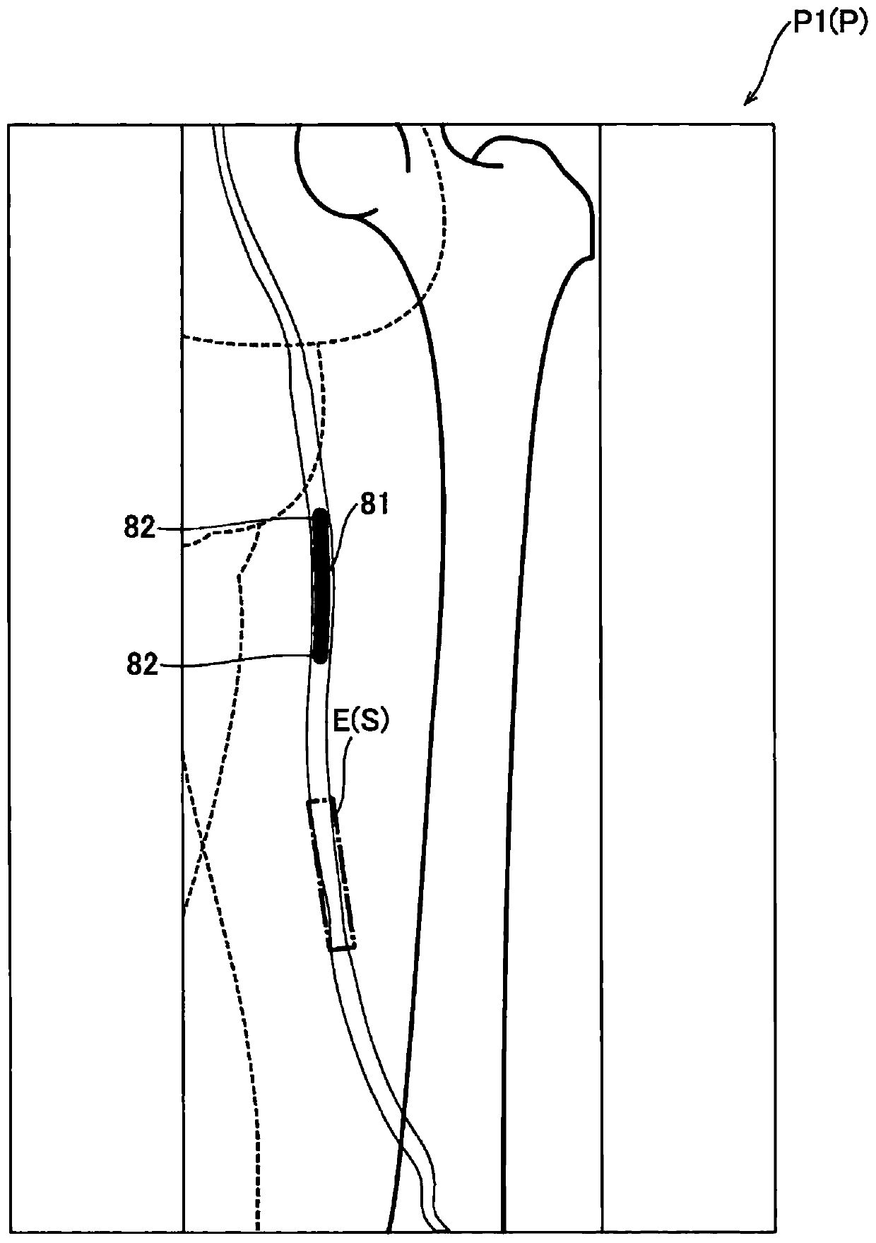
The invention provides a radiographic imaging apparatus. The radiographic imaging apparatus includes an image processor configured to perform image processing on a radiation image generated by an image generator; and a display configured to display a processed image that has been subjected to the image processing in the image processor. The image processor is configured to perform control to create the processed image showing an expanded portion in which a balloon introduced into a subject is expanded.

Description

technical field [0001] The present invention relates to a radiographic apparatus, and in particular, to a radiographic apparatus including a display unit for displaying a processed image obtained by image processing in an image processing unit. Background technique [0002] Conventionally, a radiographic apparatus is known that includes a display unit that displays a processed image obtained by image processing a radiographic image obtained by radiographing in an image processing unit. Such a radiographic apparatus is disclosed in, for example, Japanese Patent No. 5337525. [0003] The above-mentioned Patent No. 5337525 discloses an X-ray diagnostic apparatus (radiography apparatus) equipped with a display unit for displaying an X-ray image (processed image) that is displayed on the image processing unit The image obtained by image processing. Here, the image processing unit of the X-ray diagnostic apparatus of the above-mentioned Patent No. 5337525 is configured to conver...

Claims


Application Information

Patent Timeline
Patent Type & Authority Applications(China)
IPC IPC(8): A61B6/00
CPCA61B6/46A61B6/481A61B6/12A61B6/5217A61B6/504A61B6/487A61B6/463A61B2090/3966A61B90/37A61B2090/376G06T7/246G06T7/254G06T2207/10116G06T2207/30021G06T2207/30101G06V2201/034A61B34/20G06T7/73G06T7/0016A61M2025/1079A61B2034/2065A61M25/104
Inventor 吉田贵则田中文哲
Owner SHIMADZU SEISAKUSHO CO LTD
Learn More