


Quick Research
Generate reliable direction feasibility study reports for your R&D in just a few steps.

Technical Q&A
Discover and master advanced knowledge NOW. Basics, ideas, possibilities, all at once.

Find Solutions
As an expert in R&D theories, this can generate solutions to your technical problems instantly.

Evaluate Feasibility
Analyze your overall solution with one click, know your potential R&D risks in advance.

Monitor Landscape
Get weekly tech updates, stay abreast of the latest tech innovations and key insights.
Enhanced CT image tracheal wall enhancement method, system, device and medium
A technology for CT images and trachea, which is applied in image enhancement, image analysis, image data processing, etc., and can solve problems such as blurred bronchiolar wall imaging, insufficient segmentation, and incomplete three-dimensional reconstruction of the trachea
- Summary
- Abstract
- Description
- Claims
- Application Information
AI Technical Summary
Problems solved by technology
Method used
Image


Examples
Embodiment 1
[0037] Embodiment 1, this embodiment provides a method for enhancing the tracheal wall of a CT image;
[0038] A method for enhancing the tracheal wall of an enhanced CT image, comprising:
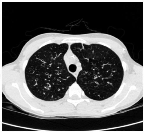
[0039] S1: Acquire an enhanced CT image sequence, and perform thresholding processing on the enhanced CT image sequence;
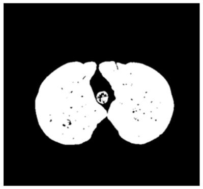
[0040] S2: Perform three-dimensional region growing on the thresholded image to obtain lung and trachea masks;
[0041] S3: Perform closed operation on the lung and trachea mask; perform three-dimensional region growth on the image obtained by the closed operation, and segment the main body area of the trachea;
[0042] S4: Remove the main body area of the trachea for each image in the enhanced CT image sequence to obtain a bronchiole image sequence; calculate the boundary pixel features of each pixel in each image in the bronchiole image sequence;
[0043] Determine whether each pixel of each image in the bronchiole image sequence belongs to the tracheal wall ba...
Embodiment 2
[0114] Embodiment 2, this embodiment also provides an enhanced CT image tracheal wall enhancement system;
[0115] An enhanced CT image tracheal wall enhancement system, comprising:
[0116] A thresholding processing module configured to: acquire a sequence of enhanced CT images, and perform thresholding processing on the sequence of enhanced CT images;
[0117] A first 3D region growing module configured to: perform 3D region growing on the thresholded image to obtain lung and trachea masks;
[0118] The second three-dimensional region growing module is configured to: perform a closed operation on the lung and trachea mask; perform three-dimensional region growth on the image obtained by the closed operation, and segment the main body area of the trachea;
[0119] An enhancement module configured to: remove the main body area of the trachea for each image in the enhanced CT image sequence to obtain a bronchiole image sequence; calculate its boundary for each pixel of eac...
Embodiment 3
[0121] Embodiment 3. This embodiment also provides an electronic device, including a memory, a processor, and computer instructions stored in the memory and run on the processor. When the computer instructions are executed by the processor, the computer instructions in Embodiment 1 are completed. steps of the method described above.
PUM
 Abstract
Description
Claims
Application Information
 - R&D Engineer
- R&D Manager
- IP Professional
- Industry Leading Data Capabilities
- Powerful AI technology
- Patent DNA Extraction
Browse by: Latest US Patents, China's latest patents, Technical Efficacy Thesaurus, Application Domain, Technology Topic, Popular Technical Reports.
© 2024 PatSnap. All rights reserved.Legal|Privacy policy|Modern Slavery Act Transparency Statement|Sitemap|About US| Contact US: help@patsnap.com