
Fundus image optic cup and optic disk segmentation method and system for assisting glaucoma screening

A fundus image and glaucoma technology, applied in image analysis, image enhancement, image data processing, etc., can solve problems such as insufficient fusion of deep and shallow features, unsatisfactory segmentation of optic cup and optic disc, insufficient semantic information level of fundus image, etc., to achieve improvement The effect of backpropagation

Active Publication Date: 2020-04-10
SICHUAN UNIV
View PDF3 Cites 35 Cited by
  • Summary
  • Abstract
  • Description
  • Claims
  • Application Information

AI Technical Summary

Problems solved by technology

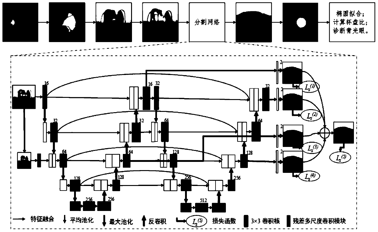
[0008] The purpose of the present invention is: in order to solve the problem that the existing network structure has insufficient levels of semantic information of fundus images and insufficient fusion of deep and shallow features, resulting in unsatisfactory segmentation of optic cup and disc. The present invention provides an auxiliary glaucoma screening The fundus image cup-optic-disc segmentation method and system can improve the accuracy of fundus image cup-optic-disc segmentation to obtain an accurate cup-to-disc ratio, thereby providing a favorable assistance for automatic glaucoma screening

Method used


Image

  • Fundus image optic cup and optic disk segmentation method and system for assisting glaucoma screening
  • Fundus image optic cup and optic disk segmentation method and system for assisting glaucoma screening
  • Fundus image optic cup and optic disk segmentation method and system for assisting glaucoma screening

Examples


Embodiment 1

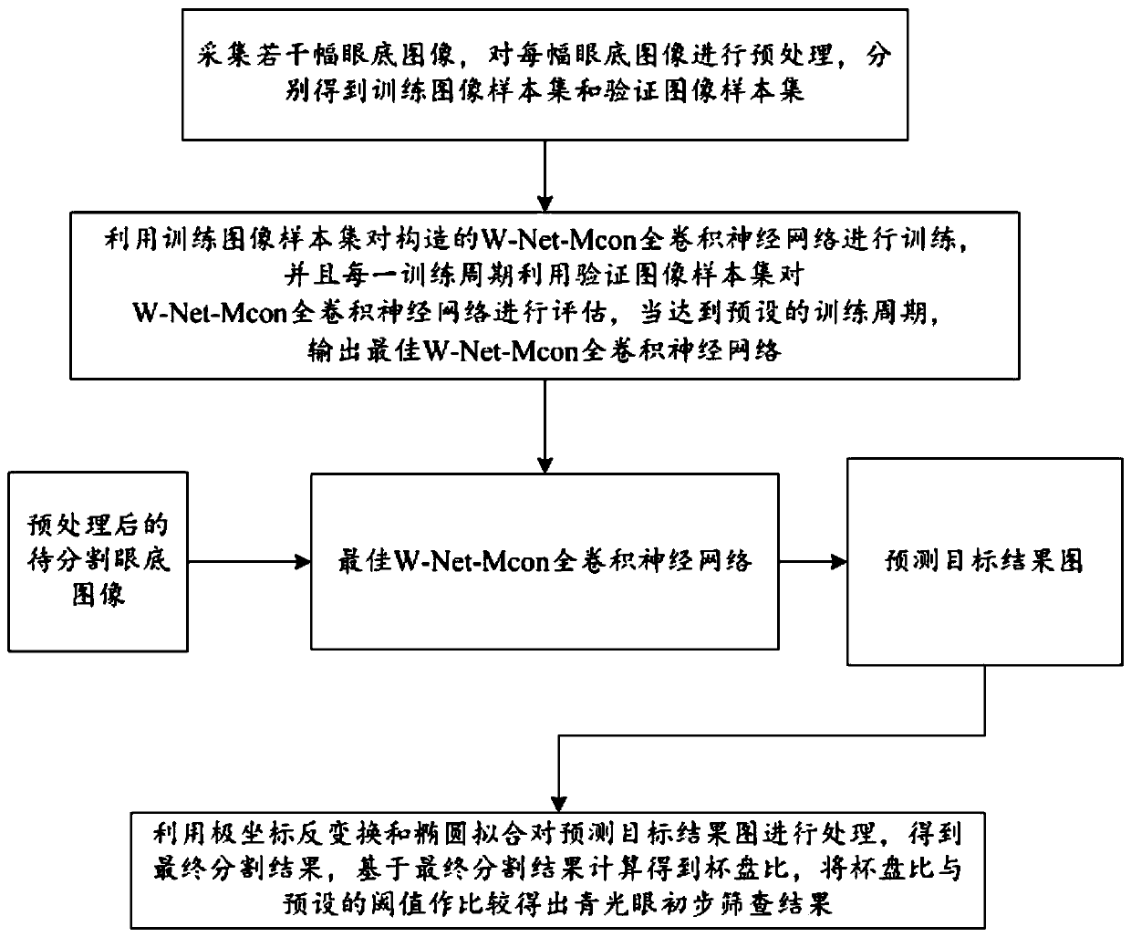
[0056] Such as figure 1 and figure 2 As shown, this embodiment provides a method for segmenting the optic cup and disc of the fundus image for assisting glaucoma screening, including the following steps:

[0057] S1: Collect several fundus images, preprocess each fundus image, and obtain training image sample sets and verification image sample sets respectively, specifically:

[0058] S1.1: Mark the two target structures of the optic disc and the optic cup in each fundus image, and obtain the corresponding target result map;

[0059] S1.2: Use the optic disc positioning method to determine the center of the optic disc in the fundus image, take the center of the optic disc as the center of interception, and intercept images of the region of interest in the same range from the fundus image and its corresponding target result map;

[0060] S1.3: Carry out polar coordinate transformation on the images of the two regions of interest respectively;

[0061] S1.4: A pair of traini...


PUM


Abstract

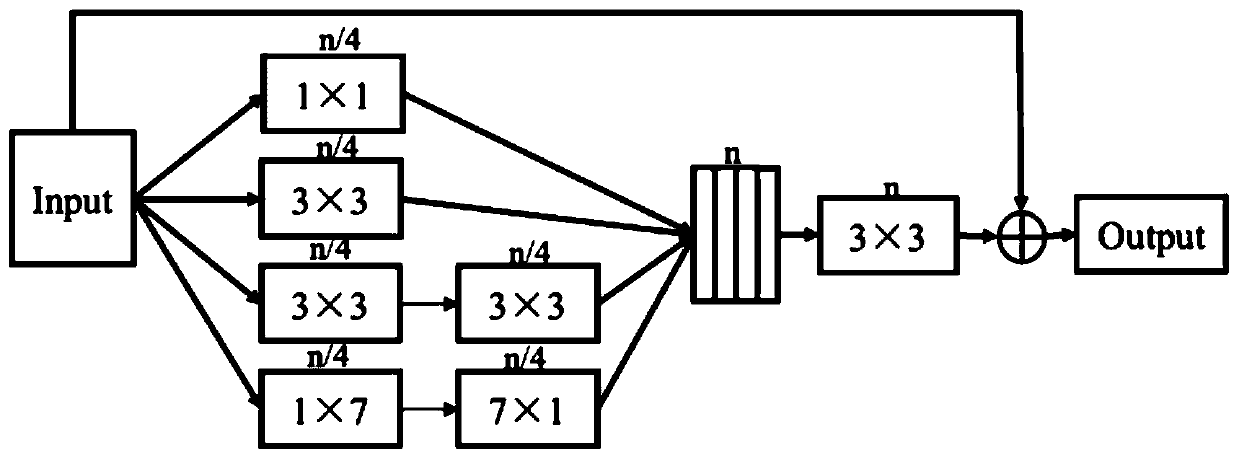
The invention discloses a fundus image optic disc segmentation method and a system for assisting glaucoma screening, and relates to the technical field of image information processing. The fundus image optic disc segmentation method comprises the steps that a plurality of fundus images are collected and preprocessed, and a training image sample set and a verification image sample set are obtained;training of a constructed W-Net-Mcon full convolutional neural network by using the training image sample set to obtain an optimal W-Net-Mcon full convolutional neural networkis carried out; preprocessing the fundus image to be segmented, and inputting the preprocessed fundus image to be segmented into the optimal W-Net-Mcon full convolutional neural network to obtain a prediction target result image; Processing prediction target result graph by utilizing polar coordinate inverse transformation and ellipse fitting to obtain final segmentation result so as to obtain cup-to-disk ratio and finally obtain glaucoma preliminary screening result. According to the method, image semantic information can be effectively extracted in a multi-size mode, fusion of features of different levels, fusion of global features and detail features and encouragement of feature multiplexing are carried out, gradient back propagation is improved, and the image segmentation precision is improved.

Description

technical field [0001] The present invention relates to the technical field of image information processing, and more specifically relates to a method and system for segmenting an optic cup and disc of a fundus image for assisting glaucoma screening. Background technique [0002] Glaucoma is the second leading cause of blindness worldwide (after cataracts) and the leading cause of irreversible blindness. Since the visual impairment caused by glaucoma is irreversible, early screening and diagnosis of glaucoma are crucial. Fundus images have become an important reference information for doctors to diagnose glaucoma due to their low cost and easy acquisition. They play a huge role in recording the process of glaucoma lesions, diagnosing glaucoma diseases, evaluating treatment effects, and preventing certain potential glaucoma lesions. . The current mainstream glaucoma screening technology is to use the optic nerve head in the fundus image to evaluate, and a binary classificat...

Claims


Application Information

Patent Timeline
Patent Type & Authority Applications(China)
IPC IPC(8): G06T7/11G06T7/00G16H50/20G06N3/08G06N3/04
CPCG06T7/11G06T7/0014G16H50/20G06N3/08G06T2207/20081G06T2207/20084G06T2207/30041G06N3/045
Inventor 郑秀娟袁鑫李淼吉彬于舒扬李彬刘凯
Owner SICHUAN UNIV
Learn More