Eureka AIR delivers breakthrough ideas for toughest innovation challenges, trusted by R&D personnel around the world.

Method for detecting post-transplantation GVHD related cytokines by applying flow cytometry and detection kit

A technology of cytokines and cytometry, applied in the field of methods and detection kits, can solve the problems of complicated means, complicated diagnostic process, and low specificity, and achieve the effects of high clinical relevance, simple method, and simple operation

Pending Publication Date: 2020-04-10
天津美瑞特医疗科技有限公司
View PDF7 Cites 8 Cited by
  • Summary
  • Abstract
  • Description
  • Claims
  • Application Information

AI Technical Summary

Problems solved by technology

The method used for the diagnosis of GVHD is biopsy, and the diagnostic process is very complicated
In view of the complexity and low specificity of the current diagnostic methods for GVHD, it is necessary to develop a rapid and specific kit for evaluating GVHD

Method used


Image

  • Method for detecting post-transplantation GVHD related cytokines by applying flow cytometry and detection kit
  • Method for detecting post-transplantation GVHD related cytokines by applying flow cytometry and detection kit
  • Method for detecting post-transplantation GVHD related cytokines by applying flow cytometry and detection kit

Examples


preparation example Construction

[0031] 1. Preparation of buffer solution

[0032] 1. Preparation of Microsphere Buffer

[0033] The microsphere buffer solution involved in this example is a PBS solution containing 5% FBS by volume, and its preparation method is: measure 50mL FBS, add 950mL pH7.2 0.1M PBS buffer solution, add 0.09%NaN 3 , mix evenly, and filter with 0.45μm water filter membrane.

[0034] 2. Preparation of sample buffer

[0035] The sample buffer solution involved in this example is a PBS solution containing 0.2% BSA (w / v), and its preparation method is: weigh 2 mg of BSA, dissolve it in 1 L of PBS buffer solution with a volume of pH 7.2 0.1M, and filter it with 0.45 μm water Membrane filtration.

[0036] 3. Preparation of Cleaning Solution

[0037] The cleaning solution involved in this embodiment is a 0.1MPBS solution with a pH of 7.2, and its preparation method is: weigh 1.482g NaH 2 PO4•2H 2 O, 14.8g Na 2 HPO 4 •12H 2 O and 8.8gNaCl, add H2O 800mL to dissolve, adjust the pH value ...

specific Embodiment approach

[0039] The preparation of all capture microspheres of the present invention is the coupling of carboxylated polystyrene microspheres with APC fluorescein and capture antibodies. The method is the same as the reaction system. Therefore, the embodiment uses the preparation of ST2 capture microspheres as follows: example. The specific implementation is as follows:

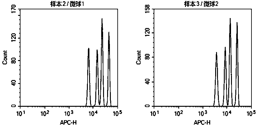
[0040] 1. Solution preparation

[0041] 1.1 Preparation of bufferA: weigh 0.96g NaH 2 PO 4 • 2H 2 O, plus H 2 O 600mL dissolved, adjust the pH value to 6.0, add H 2O was adjusted to 800mL, and filtered with a 0.45μm water filter membrane;

[0042] 1.2 Preparation of bufferB: Weigh 7.5g of glycine, add H 2 O 800mL dissolved, adjust the pH value to 7.5, add H 2 O was adjusted to 1000mL, and filtered with a 0.45μm water filter membrane;

[0043] 1.3 Preparation of 50mg / mL 1-(3-dimethylaminopropyl)-3-ethylcarbodiimide hydrochloride (EDC) solution: Weigh 1g of EDC, add 50mL of H 2 O is dissolved and filtered with...


PUM


Abstract

The invention provides a method for detecting post-transplantation GVHD related cytokines by using flow cytometry and a detection kit. The method and the detection kit have important clinical significance in the aspects of early evaluation and diagnosis of post-transplantation GVHD. According to the detection method and the kit, indexes of one or more different combinations of cell factors IL-6, IL-8, ST2, Reg3[alpha], Elafin, TNFR1, soluble IL-2R and HGF related to GVHD diagnosis and prognosis can be rapidly and homogeneously detected at the same time; single-sample multiple detection is achieved, time and resources are saved, and the result is automatically analyzed by software so as to be more objective and accurate. The method is simple, convenient, safe in detection and small in sample use quantity. By the application of the method, the risk of the GVHD of the transplanted patient can be evaluated timely, quickly and stably for a long time, the accuracy and timeliness of treatmentof the patient are improved, and the life of the patient is saved.

Description

technical field [0001] The invention belongs to the field of immunodiagnosis and detection, and in particular relates to a method and a detection kit for detecting GVHD-related cytokines after transplantation by flow cytometry. Background technique [0002] With the development of medical technology, transplantation medicine has been widely used in the treatment of various major diseases. For example: organ transplantation, bone marrow transplantation and cell transplantation, etc. However, in transplantation treatment, the postoperative complication GVHD (graft-versus-host reaction) with a postoperative morbidity rate as high as 50%-80% has always been a difficult problem for doctors. GVHD, defined as a reaction between immunocompetent cells in the graft tissue and tissues of the immunosuppressed, tissue-immiscible antigen recipient, is the leading cause of death in post-transplant patients. Therefore, evaluation, monitoring and suppression of GVHD after transplantation b...

Claims


Application Information

Patent Timeline
IPC IPC(8): G01N33/68G01N33/543G01N15/14
CPCG01N33/6863G01N33/6869G01N33/6893G01N33/54313G01N15/1434G01N2800/245G01N2800/60G01N2800/52G01N2015/1006G01N15/01
Inventor 洪建
Owner 天津美瑞特医疗科技有限公司
Why Eureka
Eureka Blog
Learn More