
Automatic liver tumor classification method and device based on physiological indexes and image fusion

A physiological index, image fusion technology, applied in the field of medical image processing, can solve the problems of improving the recognition rate, affecting the recognition performance, different tumor sizes, shapes, and positions, and achieving good robustness, automatic feature learning and extraction. , the effect of improving the recognition accuracy

Pending Publication Date: 2020-03-24
BEIJING INSTITUTE OF TECHNOLOGYGY
View PDF14 Cites 5 Cited by
  • Summary
  • Abstract
  • Description
  • Claims
  • Application Information

AI Technical Summary

Problems solved by technology

[0006] 1. The size, shape, and location of tumors in different patients are different, which brings challenges to traditional identification methods
[0007] 2. The recognition method based on traditional machine learning needs to manually design feature extraction methods for different types of liver cancer. The quality of the method design directly affects the final recognition performance
[0008] 3. The existing deep network recognition models are all developed based on image data. Such data can reflect limited tumor characteristic information, and the patient's physiological indicators have not been comprehensively used to improve the recognition rate.

Method used


Image

  • Automatic liver tumor classification method and device based on physiological indexes and image fusion
  • Automatic liver tumor classification method and device based on physiological indexes and image fusion
  • Automatic liver tumor classification method and device based on physiological indexes and image fusion

Examples


Embodiment Construction

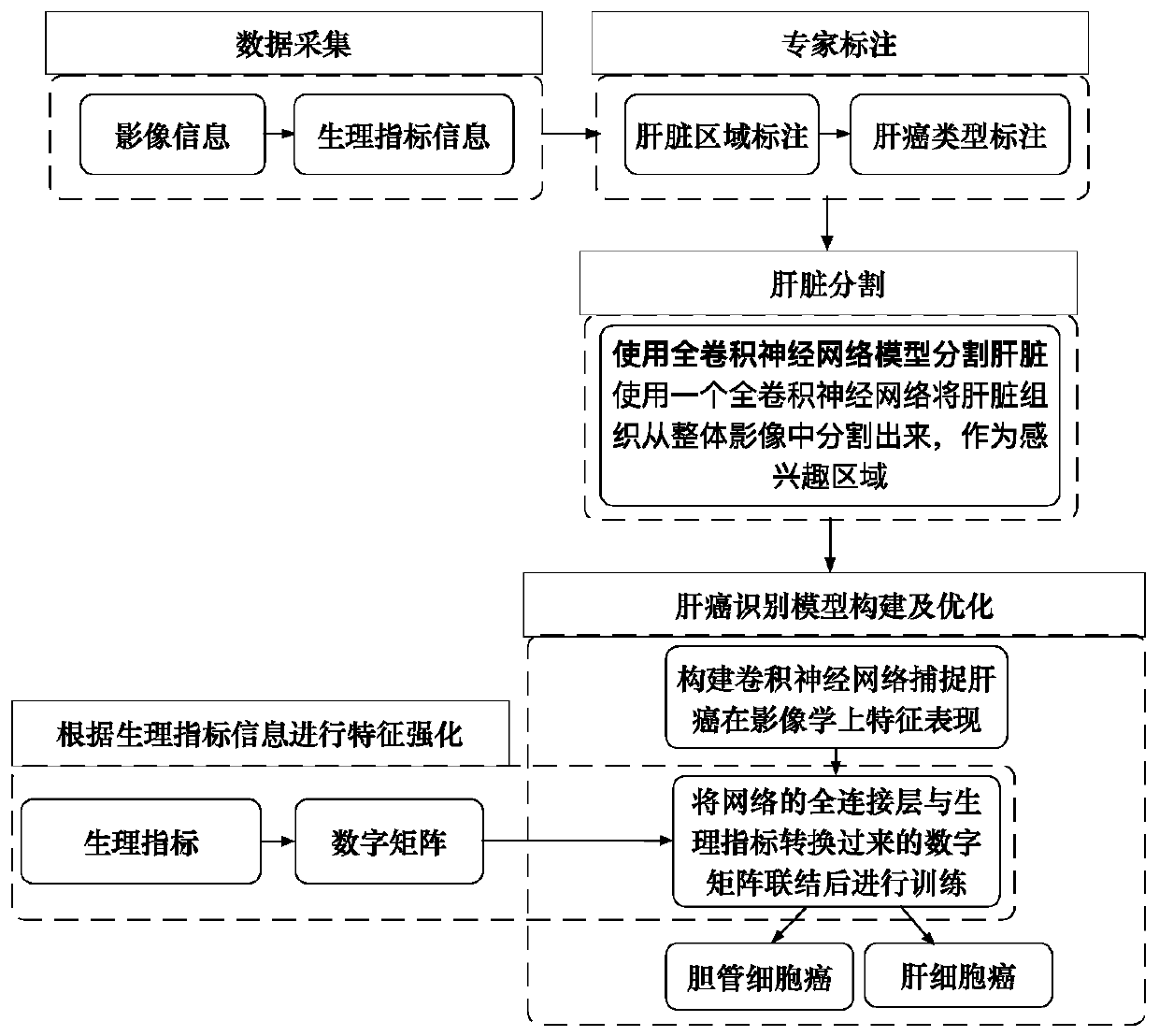
[0025] Such as figure 1 As shown, this automatic classification method for liver tumors based on physiological indicators and image fusion includes the following steps:

[0026] (1) Build a database of images and physiological indicators of cholangiocarcinoma and hepatocellular carcinoma, collect abdominal CT images of patients and corresponding physiological indicators recorded by doctors;

[0027] (2) Label all the collected image data, outline the liver tissue area and judge whether it belongs to cholangiocarcinoma or hepatocellular carcinoma, and mark it well, as the gold standard for network training;

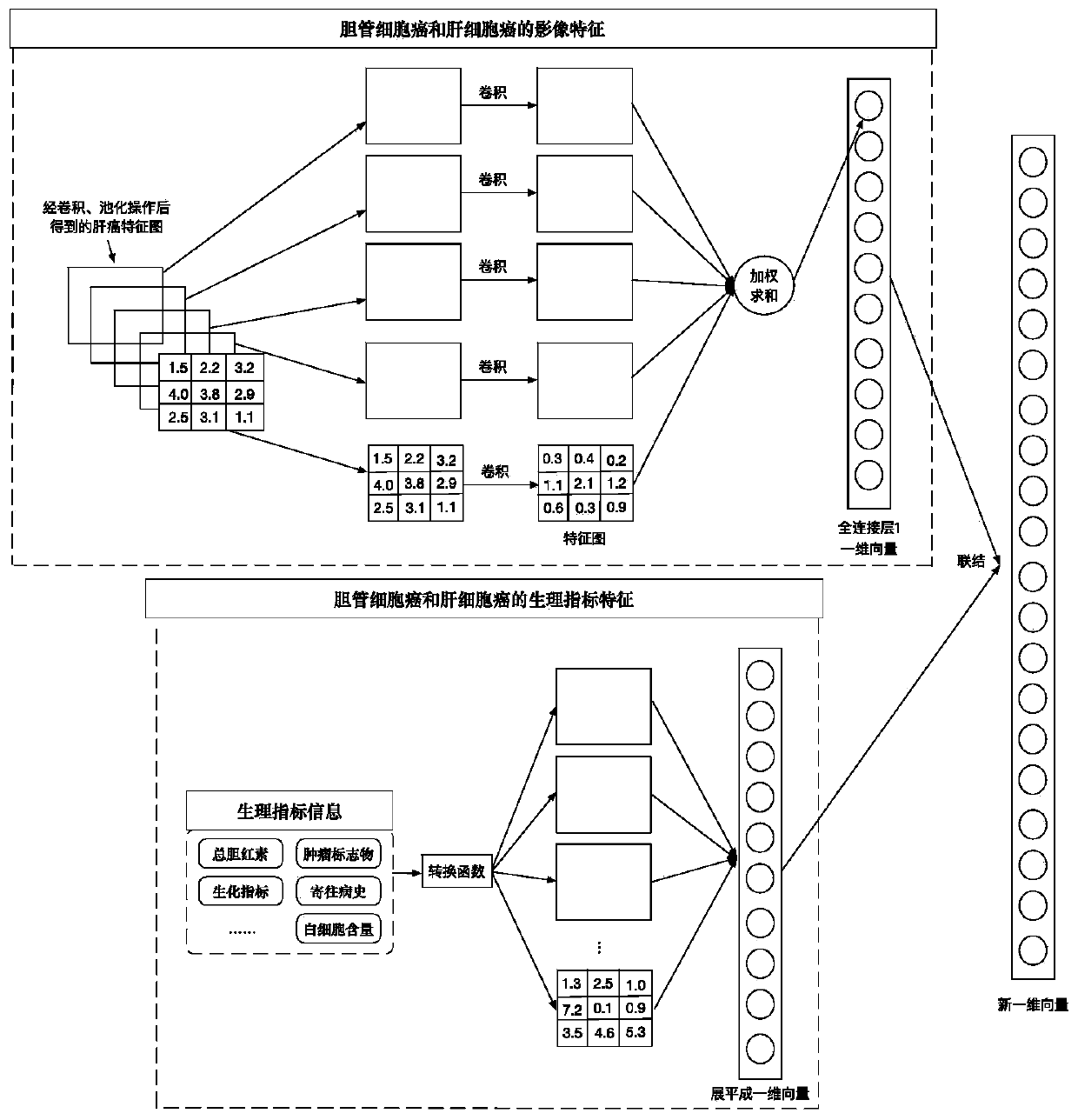
[0028] (3) Construct a three-dimensional fully convolutional neural network segmentation model, and use the image data of cholangiocarcinoma and hepatocellular carcinoma marked in the liver area in step (2) as the input of the model for learning, so that the model can automatically learn and extract liver tissue features, so that it can be segmented from the entire abdomi...


PUM


Abstract

The invention discloses an automatic liver tumor classification method and device based on physiological indexes and image fusion so that good robustness is realized for different patients during identification. A complex feature extraction algorithm does not need to be artificially designed . Full-automatic feature learning and extraction are realized, combined learning and mining are carried out on feature expression differences of the bile duct cell carcinoma and the hepatocellular carcinoma on images and expression differences of the bile duct cell carcinoma and the hepatocellular carcinoma on physiological indexes, and identification accuracy of the model is improved. The method comprises the following steps: constructing an image and physiological index database of bile duct cell carcinoma and hepatocellular carcinoma, and collecting an abdomen CT image of a patient and corresponding physiological indexes recorded by a doctor; marking all the acquired image data, drawing a livertissue area in the image data, judging whether the liver tissue area belongs to the cholangiocarcinoma or the hepatocarcinoma, and marking the liver tissue area as a gold standard for network training; constructing a three-dimensional full convolutional neural network segmentation model; and constructing a deep convolutional neural network classification model based on image and physiological index fusion.

Description

technical field [0001] The present invention relates to the technical field of medical image processing, in particular to a method for automatic classification of liver tumors based on physiological indicators and image fusion, and an automatic classification device for liver tumors based on physiological indicators and image fusion, mainly applicable to liver cancer based on computer-aided diagnosis Identify areas of study. Background technique [0002] In recent years, the incidence of cholangiocarcinoma in my country has been increasing year by year. Cholangiocarcinoma belongs to liver cancer. Its clinical manifestations are very similar to hepatocellular carcinoma. It is often misdiagnosed as hepatocellular carcinoma in clinical diagnosis, but the treatment strategies for both are different. Therefore, many patients with cholangiocarcinoma received surgery for hepatocellular carcinoma, which did not achieve the expected results and wasted a lot of medical resources. In a...

Claims


Application Information

Patent Timeline
IPC IPC(8): G06T7/00G06T7/11G06T5/50G06K9/62
CPCG06T7/0012G06T7/11G06T5/50G06T2207/10081G06T2207/20081G06T2207/20084G06T2207/20221G06T2207/30056G06T2207/30096G06F18/24
Inventor 宋红陈磊杨健艾丹妮范敬凡
Owner BEIJING INSTITUTE OF TECHNOLOGYGY
Learn More