
Device for angiocardiography

A cardiovascular and catheter technology, applied in the field of cardiovascular imaging devices, can solve the problems of increasing the degree of adhesion of contrast agents, slow injection work, long injection time, etc., to achieve the effects of easy temperature control, avoidance of excessive temperature, and strong practicability

Inactive Publication Date: 2020-02-21
THE FIRST AFFILIATED HOSPITAL OF HENAN UNIV OF SCI & TECH
View PDF0 Cites 1 Cited by
  • Summary
  • Abstract
  • Description
  • Claims
  • Application Information

AI Technical Summary

Problems solved by technology

[0002] In the basic operation of the current radiography operation, the catheter is inserted into the patient's blood vessels and corresponding body chambers, and then the contrast agent is injected into the radiography work. The device currently used is a syringe, but the viscosity of the contrast agent is extremely high. It is very laborious to inject the push-type syringe. In addition, during the imaging process, the injection work needs to be carried out slowly, otherwise it will cause danger. During the injection, the contrast agent sticks together and cannot be spread out quickly when it enters the corresponding chamber. The time for follow-up inspection is increased, and the current push-and-push injection method has major defects
In addition, when the weather is relatively cold, the degree of adhesion of the contrast agent is increased, and it is more difficult to inject such as in the patient's body.
[0003] Most of the angiography methods currently on the market are manual syringe angiography. Because it is manual injection, because each person's strength is different, the injection flow rate is also different during the injection process. Due to the high density of the contrast agent itself , is a viscous liquid, the injection flow rate is relatively slow, and the entire injection time is also long, and at the same time, the flow rate cannot be maintained at a uniform and stable level; and most of the traditional devices of this type do not have the function of contrast agent heating. It is easy to make patients feel uncomfortable, increase tension, bring inconvenience to the work of medical staff, and is not conducive to internal cleaning after use

Method used


Image

  • Device for angiocardiography
  • Device for angiocardiography
  • Device for angiocardiography

Examples


Embodiment 1

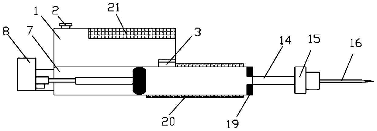
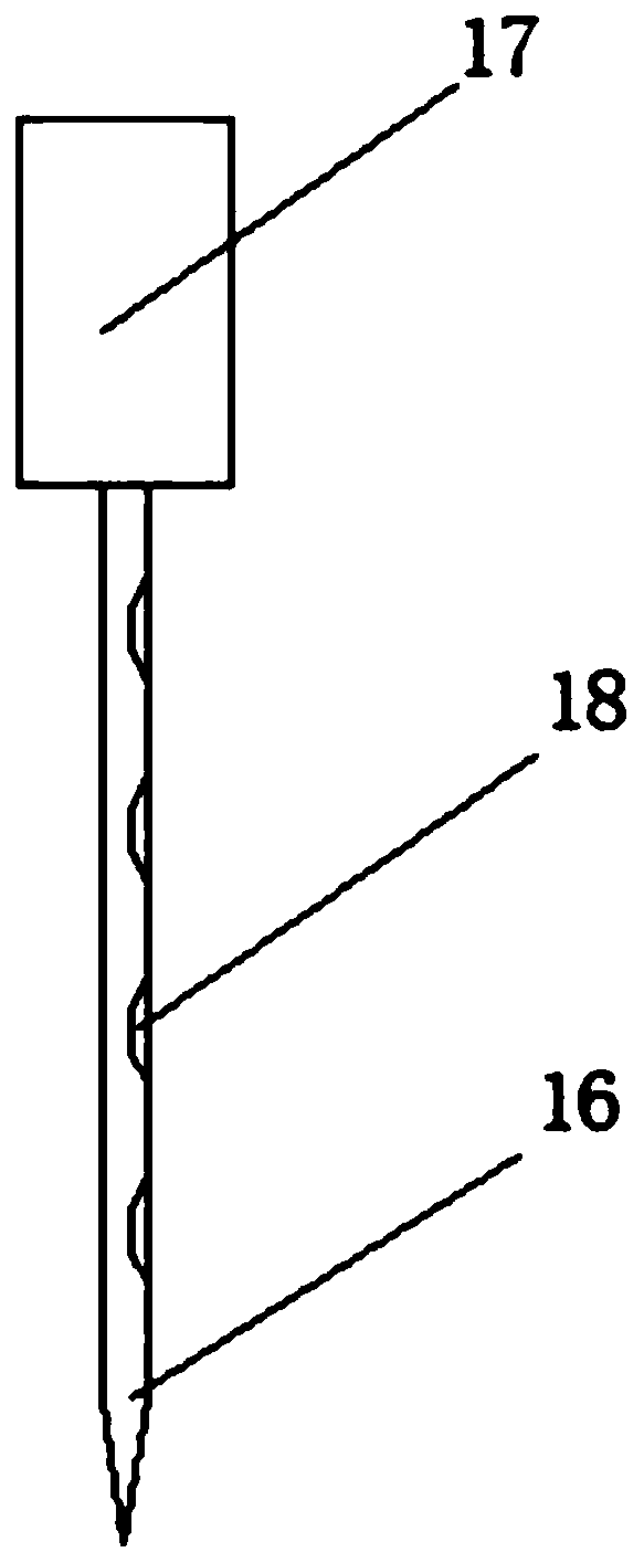
[0022] Embodiment one: Figure 1-Figure 4 A cardiovascular imaging device is shown, which includes a liquid storage box 1, a closed notch 3, a driving motor 8, and a side hole needle 16, and the top of the liquid storage box 1 is provided with a liquid injection port 2 , the top surface of the liquid storage box 1 is provided with a control panel 21, the right side of the liquid storage box 1 is provided with a closed notch 3, and the inside of the closed notch 3 is provided with a closed push plate 4, and the liquid storage box 1 is provided with a closed push plate 4. The bottom of box 1 is provided with propulsion mechanism 7, and described propulsion mechanism 7 comprises drive motor 8, electric push rod 9, main push rod 10, piston head 11 and propulsion chamber 12, and the side wall of described propulsion chamber 12 is provided with The heating block 20 electrically connected to the control panel 21, the bottom of the infusion catheter 14 is provided with a side hole nee...


PUM


Abstract

The invention discloses a device for angiocardiography. The device for the angiocardiography comprises a liquid storage box, a closed tank opening, a driving motor and a side hole needle, a liquid injection opening is arranged in the top of the liquid storage box, and the top surface of the liquid storage box is provided with a control panel; the right side of the liquid storage box is provided with the closed tank opening, and the interior of the closed tank opening is provided with a closed push plate; the bottom of the liquid storage box is provided with a propulsion mechanism, the propulsion mechanism includes the driving motor, an electric push rod, a main push rod, a piston head and a propulsion cavity, and the side wall of the propulsion cavity is provided with a heating block electrically connected to the control panel; and the bottom of a liquid conveying guide pipe is provided with the side hole needle, the right side of the propulsion cavity is provided with the liquid conveying guide pipe, and liquid leaking holes are arranged in the outer surface of the side hole needle. The device for the angiocardiography provided by the invention has a simple structure and is convenient to operate.

Description

technical field [0001] The invention relates to the field of cardiovascular medicine, in particular to a cardiovascular imaging device. Background technique [0002] In the basic operation of the current angiography operation, the catheter is inserted into the patient's blood vessels and corresponding body chambers, and then the contrast agent is injected into the angiography work. The current device used is a syringe, but the viscosity of the contrast agent is extremely high. It is very laborious to inject the push-type syringe. In addition, during the imaging process, the injection work needs to be carried out slowly, otherwise it will cause danger. During the injection, the contrast agent sticks together and cannot be spread out quickly when it enters the corresponding chamber. The time for follow-up inspection is increased, and the current push-and-push injection method has relatively large defects. In addition, when the weather is relatively cold, the degree of adhesio...

Claims


Application Information

Patent Timeline
Patent Type & Authority Applications(China)
IPC IPC(8): A61M5/20A61M5/31A61M5/32A61M5/44
CPCA61M5/007A61M5/20A61M5/31A61M5/32A61M5/445A61M2005/2006A61M2205/3368A61M2205/36
Inventor 段洪强祁娜张辉锋
Owner THE FIRST AFFILIATED HOSPITAL OF HENAN UNIV OF SCI & TECH
Learn More