
Three-dimensional reconstruction method and system based on medical image data

A technology of image data and three-dimensional reconstruction, which is applied to the details of 3D image data, image data processing, 3D modeling, etc. It can solve the problems of surgical positioning accuracy error, high price, and high misdiagnosis rate of lesions.

Pending Publication Date: 2020-01-17
上海昊骇信息科技有限公司
View PDF5 Cites 2 Cited by
  • Summary
  • Abstract
  • Description
  • Claims
  • Application Information

AI Technical Summary

Problems solved by technology

However, these medical imaging devices can only provide two-dimensional images of internal tissue or organ sections of the human body, and cannot provide continuous three-dimensional images
In the current medical diagnosis, lesions are mainly found by observing a group of CT and MRI two-dimensional slice images, and the shape and size of lesions can only be estimated by the doctor's experience in film reading, which lacks intuitiveness and is difficult to achieve accurate judgments
[0003] Traditional two-dimensional image data is difficult to read, and the display effect of the image is affected by the thickness of different machine scanning layers. Two-dimensional image data cannot give intuitive information, and the rate of misdiagnosis is high. It takes a certain amount of time for trained doctors to analyze image data. Diagnosis, and give an imaging diagnosis report, the length of the doctor's practice experience will also affect the accuracy of diagnosis; at the same time, there are large errors in the positioning accuracy of lesion surgery using two-dimensional imaging data; traditional imaging data lacks interactivity and intuition, The amount of information that can be provided for the situation that needs to calculate the three-dimensional space coordinate information is not enough
[0004] In addition, image navigation for image data positioning also exists in the existing technology, but it is expensive, and only a few hospitals have the ability to purchase related equipment, and the cost of using image equipment is also relatively high
[0005] Existing image diagnosis methods do not support remote collaborative diagnosis (equal medical treatment) or cannot perform surgical planning based on image data at the same time for multiple people

Method used


Image

  • Three-dimensional reconstruction method and system based on medical image data
  • Three-dimensional reconstruction method and system based on medical image data
  • Three-dimensional reconstruction method and system based on medical image data

Examples


Embodiment Construction

[0049] The following will clearly and completely describe the technical solutions in the embodiments of the present invention with reference to the accompanying drawings in the embodiments of the present invention. Obviously, the described embodiments are only some, not all, embodiments of the present invention. Based on the embodiments of the present invention, all other embodiments obtained by persons of ordinary skill in the art without creative efforts fall within the protection scope of the present invention.

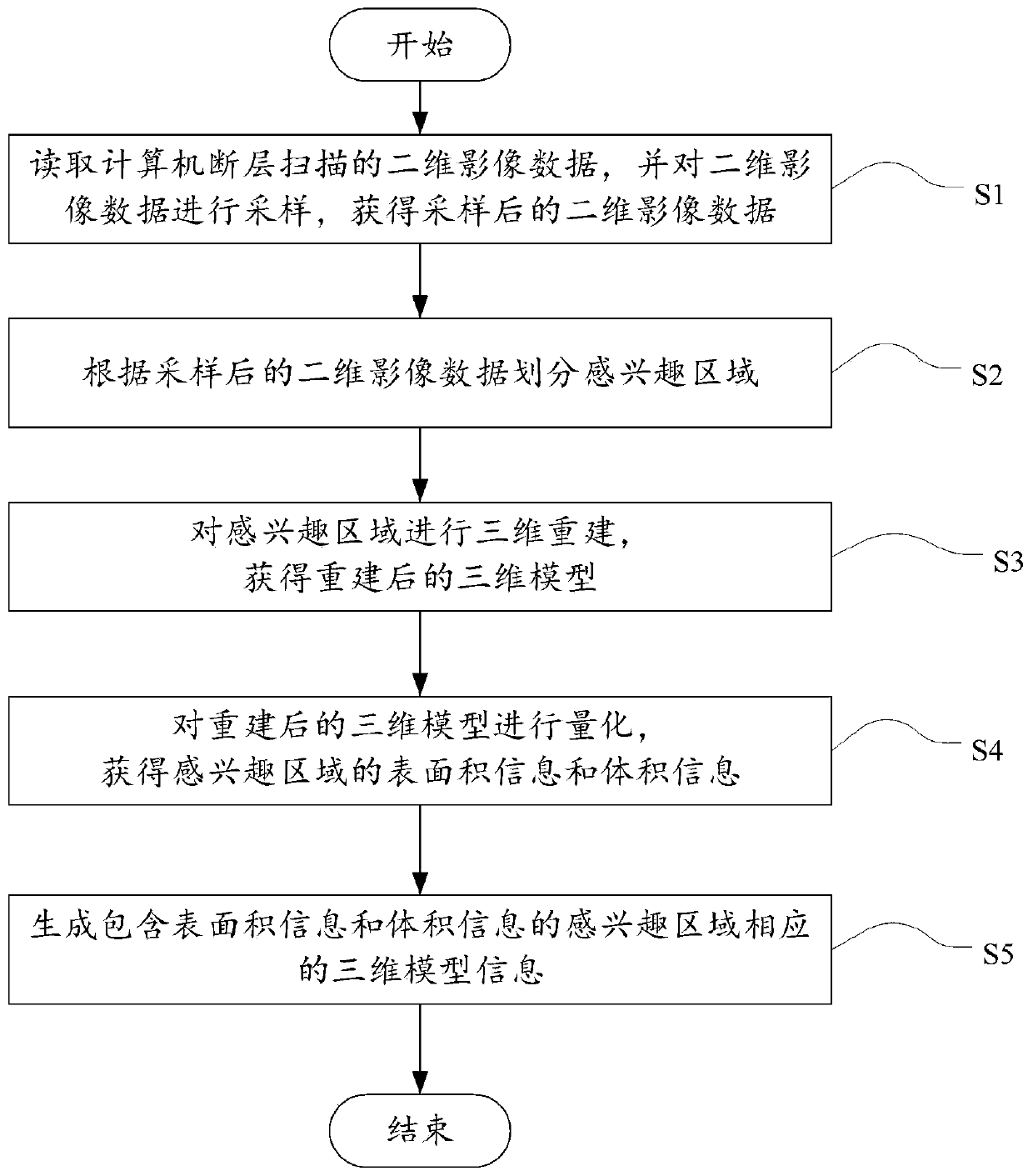
[0050] figure 1 is a schematic flow chart of a three-dimensional reconstruction method based on medical image data according to an embodiment of the present invention, such as figure 1 As shown, the method includes:

[0051] S1, reading the two-dimensional image data of computed tomography, and sampling the two-dimensional image data, and obtaining the sampled two-dimensional image data;

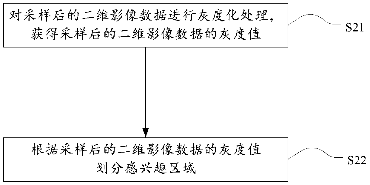
[0052] S2, dividing the region of interest according to the sampled two-di...


PUM


Abstract

The invention discloses a three-dimensional reconstruction method and system based on medical image data, and the method comprises the steps: reading two-dimensional image data of computed tomography,carrying out the sampling of the two-dimensional image data, and obtaining the sampled two-dimensional image data; dividing a region of interest according to the sampled two-dimensional image data; performing three-dimensional reconstruction on the region of interest to obtain a reconstructed three-dimensional model; quantifying the reconstructed three-dimensional model to obtain surface area information and volume information of the region of interest; and generating three-dimensional model information corresponding to the region of interest including the surface area information and the volume information. According to the embodiment of the invention, three-dimensional reconstruction is carried out on the traditional medical image data to obtain the three-dimensional image of the medical image data, more visual image data can be provided, a more comprehensive image diagnosis mode is provided, and a powerful support is provided for application modes such as preoperative planning, intraoperative navigation, remote assistance and simulated surgery.

Description

technical field [0001] The invention relates to the technical fields of medical image processing and computer image processing, in particular to a three-dimensional reconstruction method and system based on medical image data. Background technique [0002] With the development of modern medical imaging technology, there have been computed tomography (Computed Tomography, CT), magnetic resonance imaging (Magnetic Resonance Imaging, MRI), positron emission computed tomography (Positron Emission Computed Tomography, PET), ultrasound and so on. However, these medical imaging devices can only provide two-dimensional images of internal tissues or organ sections of the human body, and cannot provide continuous three-dimensional images. In the current medical diagnosis, lesions are mainly found by observing a group of CT and MRI two-dimensional slice images, and the shape and size of lesions can only be estimated by the doctor's experience in film reading, which lacks intuitiveness ...

Claims


Application Information

Patent Timeline
Patent Type & Authority Applications(China)
IPC IPC(8): G06T17/00G06T7/33G06K9/62
CPCG06T17/00G06T7/33G06T2207/20104G06T2200/04G06F18/22
Inventor 赵一阳束旭俊杨国栋
Owner 上海昊骇信息科技有限公司
Learn More