
Dual-mode tomography method, device and system

A technology of dual-mode imaging and combination method, which is applied in the field of biomedical imaging to achieve the effect of improving signal quality and improving the effect of follow-up imaging

Active Publication Date: 2019-12-20
王雪梅 +2
View PDF8 Cites 1 Cited by
  • Summary
  • Abstract
  • Description
  • Claims
  • Application Information

AI Technical Summary

Problems solved by technology

[0005] For this reason, the purpose of the present invention is to propose a dual-mode imaging method, device and system, which can solve the technical problem that the SPECT equipment composed of photomultiplier tubes cannot operate normally in the working environment of MR equipment, and can detect It can collect signal data at the same time for imaging, improve the quality of the collected signal, and improve the follow-up imaging effect

Method used


Image

  • Dual-mode tomography method, device and system
  • Dual-mode tomography method, device and system
  • Dual-mode tomography method, device and system

Examples


Embodiment Construction

[0021] Embodiments of the present invention are described in detail below, examples of which are shown in the drawings, wherein the same or similar reference numerals designate the same or similar elements or elements having the same or similar functions throughout. The embodiments described below by referring to the figures are exemplary only for explaining the present invention and should not be construed as limiting the present invention. On the contrary, the embodiments of the present invention include all changes, modifications and equivalents coming within the spirit and scope of the appended claims.

[0022] In order to solve the low signal-to-noise ratio of MR equipment with low magnetic field in related technologies, the serial combination of SPECT equipment and MR equipment cannot realize simultaneous acquisition of signal data, and the signal quality of signal data in a low magnetic field environment Not good, affecting the technical problem of poor subsequent imagi...


PUM


Abstract

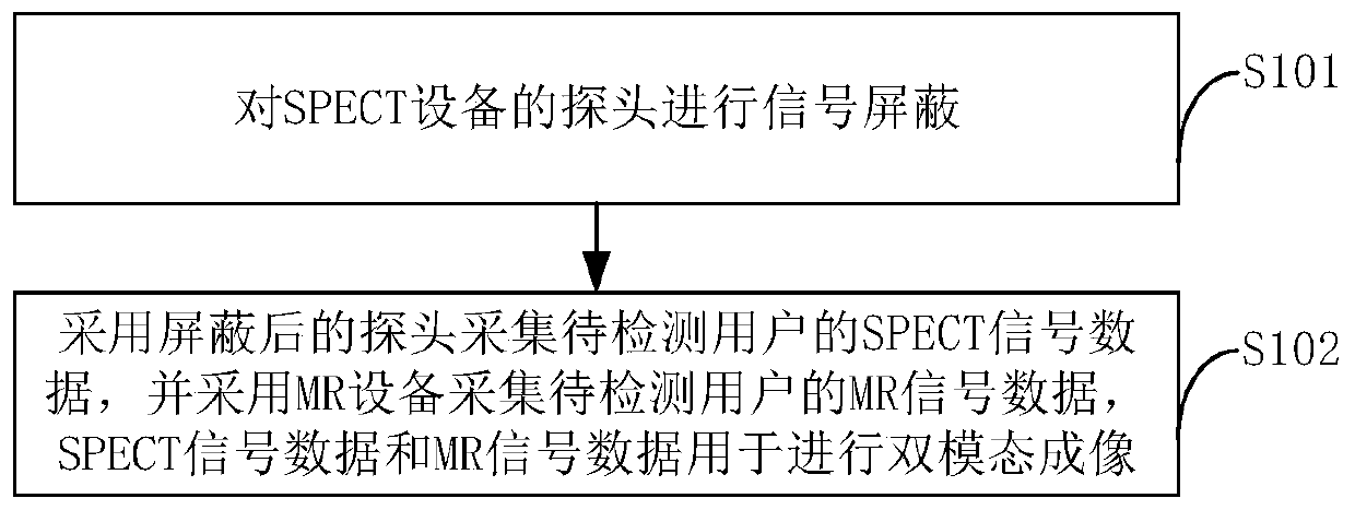
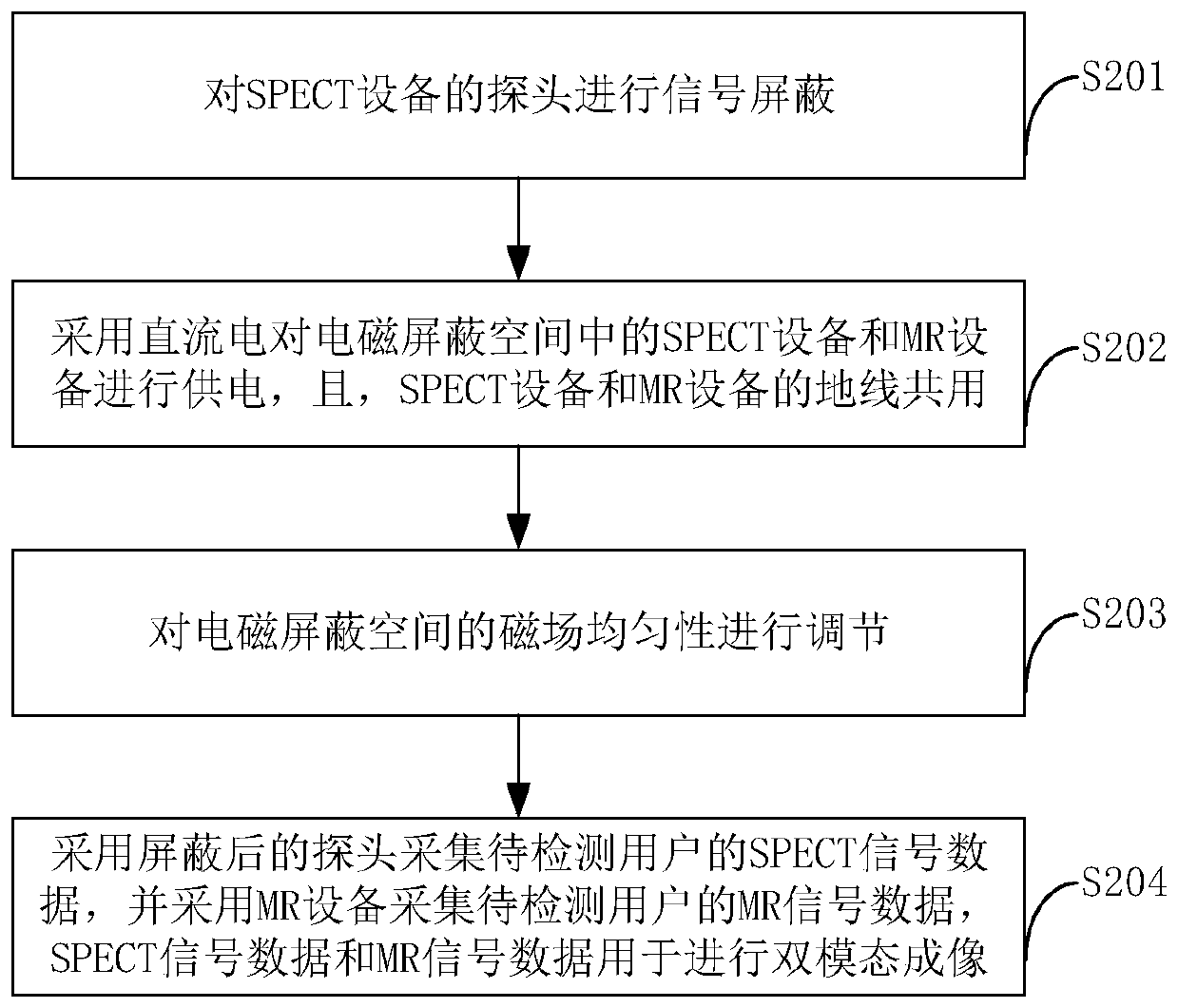
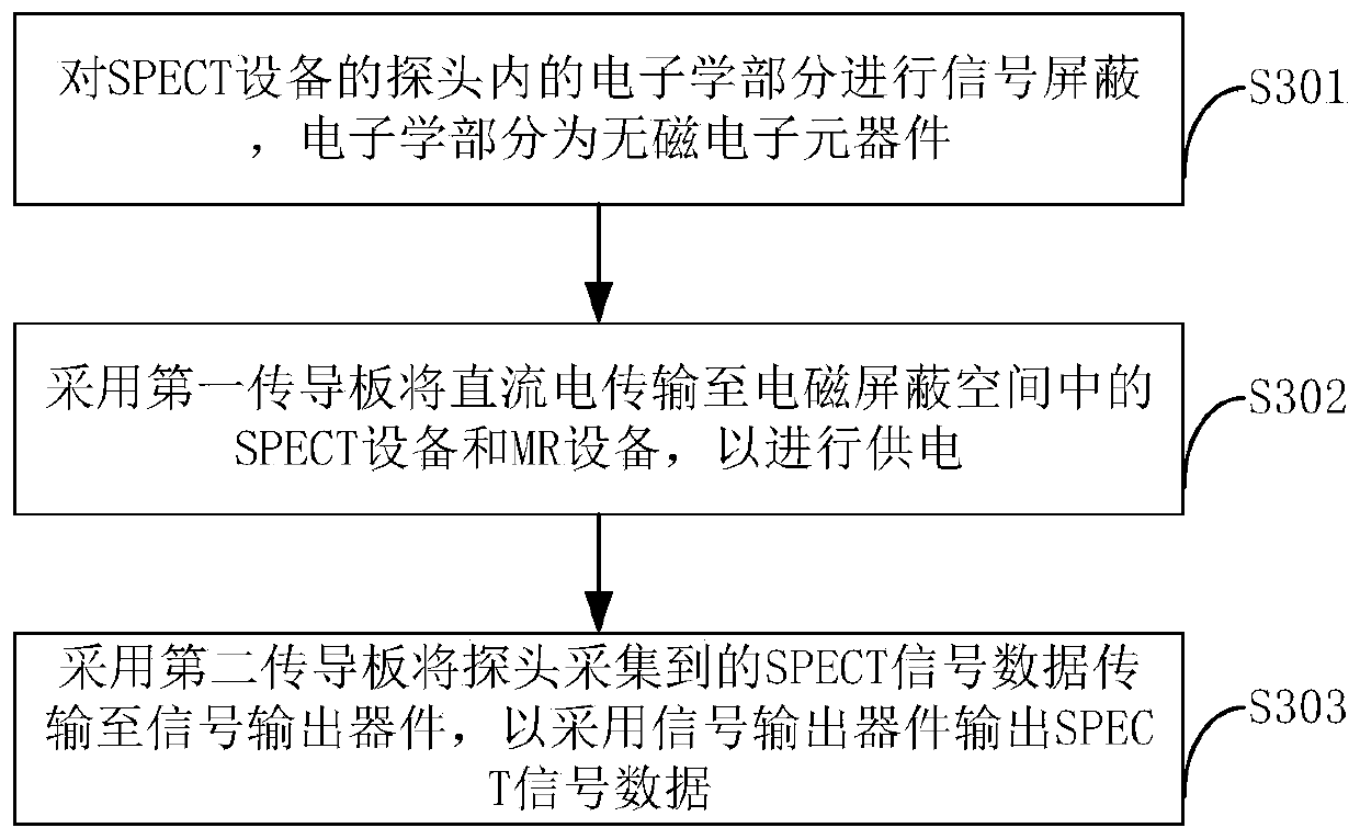
The invention provides a dual-mode tomography method, device and system which are applied to dual-mode tomography equipment. The dual-mode tomography equipment comprises SPECT equipment and MR equipment, and the SPECT equipment and the MR equipment are arranged in an electromagnetic shielding space. The method comprises the following steps of performing signal shielding on a probe of the SPECT equipment; collecting SPECT signal data of a user to be detected by the shielded probe; and collecting MR signal data of the user to be detected by the MR equipment, wherein the SPECT signal data and theMR signal data are used for performing dual-mode tomography. By using the dual-mode tomography method, device and system provided by the invention, the problem that conventional SPECT equipment of adetector formed by a photomultiplier cannot normally operate in the work environment of the MR equipment can be solved; during detection, signal data can be collected at the same time for tomography;the quality of the collected signals is improved; and the subsequent tomography effect is improved.

Description

technical field [0001] The invention relates to the technical field of biomedical imaging, in particular to a dual-mode imaging method, device and system. Background technique [0002] SPECT (Single-Photon Emission Computed Tomography, Single-Photon Emission Computed Tomography) is an imaging device for clinical nuclear medicine diagnosis, which can provide functional information at the molecular level, and has high imaging sensitivity, but its resolution is low, making it difficult to accurately describe lesions scope. MRI (Magnetic Resonance Imaging, Magnetic Resonance Imaging) is a kind of tomographic imaging, which can use the magnetic resonance phenomenon to obtain electromagnetic signals from the user to be detected, and perform spatial encoding to reconstruct the image of the user to be detected. It has high resolution and can obtain material A variety of physical characteristic parameters, such as proton density, spin-lattice relaxation time T1, etc. [0003] In th...

Claims


Application Information

Patent Timeline
IPC IPC(8): A61B6/03A61B5/055
CPCA61B6/037A61B6/482A61B6/5235A61B5/055A61B5/0035A61B2562/182
Inventor 王雪梅高丽蕾王相成包宝亮何玉林张志勇张义权吴志强银建伟刘迈王道宇侯岩松
Owner 王雪梅
Learn More