Radial golden angle magnetic resonance heart film imaging method and device and equipment
An imaging method and magnetic resonance technology, applied in the field of medical imaging, can solve the problems of slow imaging process and time-consuming, and achieve the effect of improving the quality and shortening the time of image reconstruction.
- Summary
- Abstract
- Description
- Claims
- Application Information
AI Technical Summary
Problems solved by technology
Method used
Image

Examples
Embodiment 1
[0029] figure 1 The flow chart of the radial golden angle magnetic resonance cardiac cine imaging method provided by Embodiment 1 of the present invention, this embodiment is applicable to the case of performing magnetic resonance cardiac cine imaging according to radial golden angle sampling data, the method can be performed by radial The golden angle magnetic resonance cardiac cine imaging device can be implemented specifically by software and / or hardware in electronic equipment, wherein the electronic equipment can be a magnetic resonance scanner.
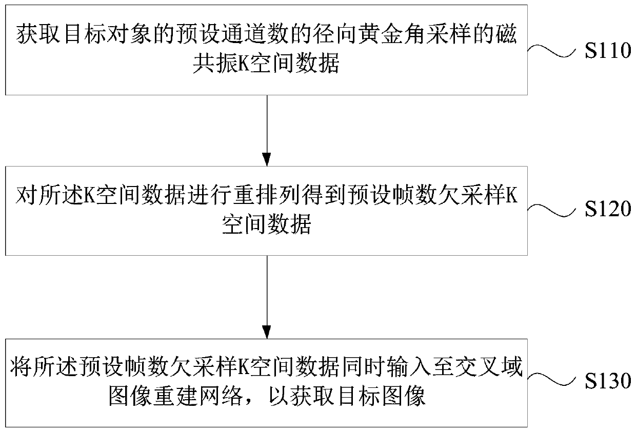
[0030] Such as figure 1 As shown, the radial golden angle magnetic resonance cardiac cine imaging method specifically includes:
[0031] S110. Obtain magnetic resonance K-space data of the radial golden angle sampling of the preset channel number of the target object.
[0032] Wherein, the target object may be a human or an animal, and when it needs to diagnose heart disease or evaluate heart function, it becomes the scanning ...
Embodiment 2
[0073] Image 6 A schematic structural diagram of a radial golden angle magnetic resonance cardiac cine imaging device provided for Embodiment 2 of the invention. This embodiment is applicable to the case of magnetic resonance cardiac cine imaging based on radial golden angle sampling data. The device can be configured in In an MRI scanner.
[0074] Such as Image 6 As shown, the radial golden angle magnetic resonance cardiac cine imaging device provided in the embodiment of the present invention includes: a data acquisition module 610 , a data preprocessing module 620 and an image reconstruction module 630 .
[0075] Among them, the data acquisition module 610 is used to acquire the magnetic resonance K-space data of the radial golden angle sampling of the preset channel number of the target object; the data preprocessing module 620 is used to rearrange the K-space data to obtain the preset The K-space data under-sampled by the number of frames; the image reconstruction mod...
Embodiment 3
[0091] Figure 7 It is a structural schematic diagram of the magnetic resonance scanning system in Embodiment 3 of the present invention, and the magnetic resonance scanning system includes: scanning equipment, a treatment couch, and computer equipment.
[0092] Among them, the scanning equipment is used to acquire magnetic resonance scanning data; the user of the treatment bed carries the target object receiving the magnetic resonance scanning, and moves the target object to the designated scanning position; the computer equipment is used to control the working process of the scanning equipment and the treatment bed, thereby After completing the magnetic resonance scan, it can also be used to obtain the magnetic resonance scan data and process the data to obtain the reconstructed target image.
[0093] Further, the schematic diagram of the hardware structure of the computer equipment can be referred to Figure 8 .
[0094] Figure 8 It is a schematic structural diagram of ...
PUM
 Abstract
Description
Claims
Application Information
 - R&D
- Intellectual Property
- Life Sciences
- Materials
- Tech Scout
- Unparalleled Data Quality
- Higher Quality Content
- 60% Fewer Hallucinations
Browse by: Latest US Patents, China's latest patents, Technical Efficacy Thesaurus, Application Domain, Technology Topic, Popular Technical Reports.
© 2025 PatSnap. All rights reserved.Legal|Privacy policy|Modern Slavery Act Transparency Statement|Sitemap|About US| Contact US: help@patsnap.com