
A sub-millisecond real-time 3D super-resolution microscopy imaging system

A real-time three-dimensional, microscopic imaging technology, applied in microscopes, analytical materials, material excitation analysis, etc., can solve problems such as inapplicability, unsuitability for rapid molecular motion process monitoring, PSF damage, etc.

Active Publication Date: 2021-06-22
FUDAN UNIV
View PDF7 Cites 0 Cited by
  • Summary
  • Abstract
  • Description
  • Claims
  • Application Information

AI Technical Summary

Problems solved by technology

[0004] However, the above methods are not suitable for the monitoring of fast molecular motion process.
First, due to the high speed of molecular motion, the PSF of the molecule will be destroyed, and all PSF-based techniques will not be applicable

Method used


Image

  • A sub-millisecond real-time 3D super-resolution microscopy imaging system
  • A sub-millisecond real-time 3D super-resolution microscopy imaging system

Examples


Embodiment Construction

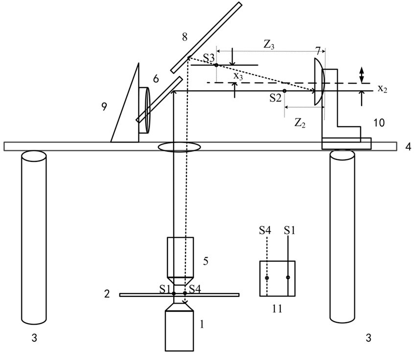
[0018] Refer figure 1 As shown, the sample is inverted the fluorescent microscope laser light source excitation, producing a fluorescent signal, and the first object mirror M1 1 of the inverted fluorescent microscope is from 0.9, and the lower hemispherical surface original mono-molecular fluorescence signal S1 is collected. The flat surface of the inverted microscope is called the first plane. On the first plane 2, the optical plate 4 is connected to the second plane by the stent 3, and the second plane must be securely securely securely fixed, and independently of the external system to achieve the purpose of reducing vibration. The first planar mirror 6, the concave mirror M3 7, the second plane 8, a triangular frame 9, and the L-shaped frame 10 are placed on the second plane 4. S1 is the original fluorescent signal, S2 is the image point after S1 passing through the second objective mirror M2 5, and S3 is the imaging point after the image point S2 passes through the concave mi...


PUM


Abstract

The invention belongs to the technical field of single-molecule imaging, and in particular relates to a submillisecond-level real-time three-dimensional super-resolution microscopic imaging system. The system of the present invention comprises: the first objective lens, the second objective lens, the first plane, the second plane, the first plane mirror, the concave mirror, the second plane mirror, and EMCCD; By building the second objective lens, the fluorescence signal on the upper hemisphere is collected to the second plane, and then returned to the sample plane by the concave mirror on the second plane; after being enlarged by the second objective lens, the single molecule signal is separated from the central axis of the concave mirror The distance is close to the focal length of the concave mirror, and the best measurement accuracy in the vertical direction is achieved.

Description

Technical field [0001] The present invention belongs to the field of single molecular imaging, and in particular to a mi-miocither real-time three-dimensional ultra-resolution micro-imaging system. Background technique [0002] Super resolution microscope brings an optical microscope into a nanoscale, achieving visualization of a single molecular passage in living cells. High-time resolution and high-dimensional spatial resolution microscope are widely used to further study the mechanism of cell nuclear pore protein regulatory genes, millisecond magnitude and even faster cilia amimini-molecular transport, cytocnectic molecular exchange and transmembrane channel switch and other biological problems. Therefore, the super resolution microscope experiment system is required to have high-time resolution and high-dimensional spatial distinguishing photoelectric signal detection function. [0003] Today, the super resolution microscope is mainly divided into two major categories. One ty...

Claims


Application Information

Patent Timeline
Patent Type & Authority Patents(China)
IPC IPC(8): G01N21/64G02B21/00G02B21/02
CPCG01N21/64G01N21/6486G01N2021/6423G02B21/0088G02B21/02
Inventor 马炯刘晓兰
Owner FUDAN UNIV
Learn More