Preparation method of in-vitro human testicular spermatogenesis model
A technology of spermatogenesis and testis, applied in the field of biomedicine, can solve problems such as unreached
- Summary
- Abstract
- Description
- Claims
- Application Information
AI Technical Summary
Problems solved by technology
Method used
Image

Examples
Embodiment 1
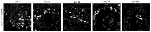
[0022] Example 1 The spermatogonial stem cells still proliferated in the testes of 12-week-embryo cultured for 100 days
[0023] (1) Tissue culture method: prepare 0.25% agar blocks with low melting point agar and sterilized water, cut the agar blocks into 8mm×8mm squares, and replace them with complete testis medium for at least 8 hours; Remove the mesonephros from the human embryonic testes, cut them into 3mm tissue pieces, place them on the prepared 0.25% agar block soaked in the culture set in advance, add the culture solution to two-thirds of the agar block, and 34°C, 5% CO 2 to cultivate;
[0024] (2) Establishment of culture system: During the cultivation process of this experiment, in order to find out the most suitable medium for testicular organogenesis in vitro, three culture schemes were adopted in the experiment. The specific medium components are as follows:
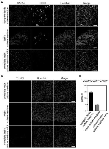
[0025]Complete testis culture medium: 90% (V / V) αMEM, 10% (V / V) KSR, BMP 4 / 7 (20 ng / mL, R&D Systems), ...
Embodiment 2
[0030] Example 2 The spermatocytes appeared in the 12-week-old testis cultured for 10 days
[0031] (1) Tissue culture method: prepare 0.25% agar blocks with low melting point agar and sterilized water, cut the agar blocks into 8mm×8mm squares, and replace them with complete testis medium for at least 8 hours; Remove the mesonephros from the human embryonic testes, cut them into 3mm tissue pieces, place them on the prepared 0.25% agar block soaked in the culture set in advance, add the culture solution to two-thirds of the agar block, and 34°C, 5% CO 2 to cultivate;
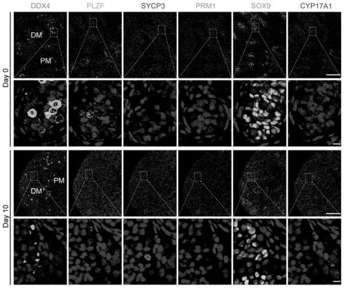
[0032] (2) Using complete testis medium to culture 12-week human testis for 10 days, stain the tissue sections from Day 0 before culture and Day 10 after culture, the cell types that play a key role in spermatogenesis, including germ cells, spermatogonia, and spermatozoa Cell markers (DDX4, PLZF, SYCP3, PRM1, SOX9, CYP17A1) of sperm cells, Sertoli cells, and mesenchymal cells were fluorescently stained ( imag...
Embodiment 3
[0033] Example 3 Round spermatozoa appeared in the 12-week-embryo testes after 30 days of culture
[0034] (1) Tissue culture method: prepare 0.25% agar blocks with low melting point agar and sterilized water, cut the agar blocks into 8mm×8mm squares, and replace them with complete testis medium for at least 8 hours; Remove the mesonephros from the human embryonic testes, cut them into 3mm tissue pieces, place them on the prepared 0.25% agar block soaked in the culture set in advance, add the culture solution to two-thirds of the agar block, and 34°C, 5% CO 2 to cultivate;
[0035] (2) The 12-week human testis was cultured for 30 days with complete testis medium, and the tissue sections at 30 days during the culture process were stained, and markers for germ cells, spermatogonia, spermatocytes, sperm cells, Sertoli cells, and mesenchymal cells ( DDX4, PLZF, SYCP3, PRM1, SOX9, CYP17A1) for fluorescent staining ( Figure 4 ), the results showed that there were positive signal...
PUM
| Property | Measurement | Unit |
|---|---|---|
| diameter | aaaaa | aaaaa |
Abstract
Description
Claims
Application Information
 - R&D
- Intellectual Property
- Life Sciences
- Materials
- Tech Scout
- Unparalleled Data Quality
- Higher Quality Content
- 60% Fewer Hallucinations
Browse by: Latest US Patents, China's latest patents, Technical Efficacy Thesaurus, Application Domain, Technology Topic, Popular Technical Reports.
© 2025 PatSnap. All rights reserved.Legal|Privacy policy|Modern Slavery Act Transparency Statement|Sitemap|About US| Contact US: help@patsnap.com