Blood vessel image processing method, device and apparatus and storage medium
A blood vessel image and processing method technology, applied in the field of blood vessel image processing method, device, equipment and storage medium, can solve the problems affecting the reliability of diagnosis results and the like
- Summary
- Abstract
- Description
- Claims
- Application Information
AI Technical Summary
Problems solved by technology
Method used
Image

Examples
Embodiment 1
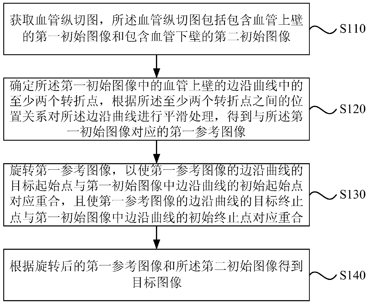
[0038] Figure 1A It is a flowchart of a blood vessel image processing method in Embodiment 1 of the present invention. The embodiment of the present invention is applicable to the situation of removing jitter noise caused by respiration and heartbeat in the collected ultrasound images of blood vessels. The method is performed by a blood vessel image processing device, which is implemented by software and / or hardware, and is specifically configured in an electronic device capable of image processing, and the electronic device may be an independent computing device, such as a personal computer or a PC, etc. , may also be a data processing device contained in a medical imaging system, where the medical imaging device may be an ultrasonic diagnostic device.
[0039] Such as Figure 1A A blood vessel image processing method shown includes:
[0040] S110. Acquire a longitudinal sectional image of a blood vessel, where the longitudinal sectional image of a blood vessel includes a f...
Embodiment 2
[0056] Figure 2A It is a flowchart of a blood vessel image processing method in Embodiment 2 of the present invention. The embodiments of the present invention are optimized and improved on the basis of the technical solutions of the foregoing embodiments.
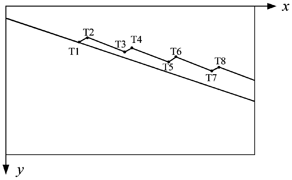
[0057] Further, the operation "smoothing the edge curve according to the positional relationship between the at least two turning points" is refined as "dividing the edge curve into at least three The initial curve segmentation segment; according to the overall direction of the blood vessel, at least two target curve segmentation segments are selected from each of the initial curve segmentation segments; two adjacent endpoints of the two adjacent target curve segmentation segments are grouped Turning point pairs, according to each group of turning point pairs, the target curve segment is translated, so that the at least two target curve segment segments are "collinear", so as to improve the processing method of smoothing...
Embodiment 3
[0078] image 3 It is a flowchart of a blood vessel image processing method in Embodiment 3 of the present invention. The embodiments of the present invention are optimized and improved on the basis of the technical solutions of the foregoing embodiments.
[0079] Further, the operation "rotate the first reference image, so that the target starting point of the edge curve in the first reference image coincides with the initial starting point of the edge curve in the first initial image, and make the The target end point of the edge curve of the first reference image coincides with the initial end point of the edge curve in the first initial image correspondingly "refinement" to "surround the first reference image around the target start point or the The target end point is rotated so that the target start point coincides with the initial start point, and the target end point coincides with the initial end point", so as to improve the rotation of the first reference image. ro...
PUM
 Abstract
Description
Claims
Application Information
 - R&D
- Intellectual Property
- Life Sciences
- Materials
- Tech Scout
- Unparalleled Data Quality
- Higher Quality Content
- 60% Fewer Hallucinations
Browse by: Latest US Patents, China's latest patents, Technical Efficacy Thesaurus, Application Domain, Technology Topic, Popular Technical Reports.
© 2025 PatSnap. All rights reserved.Legal|Privacy policy|Modern Slavery Act Transparency Statement|Sitemap|About US| Contact US: help@patsnap.com